
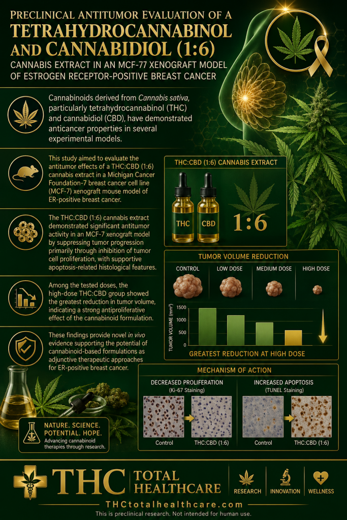
“Background and aim: Breast cancer remains one of the leading causes of cancer-related mortality worldwide, despite advances in surgery, chemotherapy, endocrine therapy, and targeted treatments. Cannabinoids derived from Cannabis sativa, particularly tetrahydrocannabinol (THC) and cannabidiol (CBD), have demonstrated anticancer properties in several experimental models; however, in vivo evidence in estrogen receptor (ER)-positive breast cancer remains limited. This study aimed to evaluate the antitumor effects of a THC:CBD (1:6) cannabis extract in a Michigan Cancer Foundation-7 breast cancer cell line (MCF-7) xenograft mouse model of ER-positive breast cancer.
Materials and methods: Female BALB/c nude mice bearing MCF-7 xenograft tumors were randomly assigned into five groups (n = 5/group): negative control (sesame oil), positive control treated with 5-fluorouracil (5-FU; 20 mg/kg), and three treatment groups receiving oral THC:CBD (1:6) extract at doses of 2, 10, or 20 mg/kg body weight for 30 consecutive days. Tumor growth was monitored throughout the experiment. Histopathological examination and immunohistochemical analysis of proliferating cell nuclear antigen (PCNA) expression were performed to evaluate apoptosis-related morphology and tumor cell proliferation. Hematological and biochemical parameters were assessed to determine systemic safety.
Results: Cannabinoid-treated groups exhibited significant suppression of tumor growth compared with the negative control group. Tumor volume reduction was observed in all treatment groups, with the greatest reduction detected in the high-dose THC:CBD group. Histopathological evaluation revealed increased numbers of tumor cells exhibiting morphological features consistent with apoptosis in cannabinoid-treated mice. Immunohistochemical analysis demonstrated significantly lower PCNA expression scores in all THC:CBD-treated groups compared with both negative and positive controls, indicating reduced tumor cell proliferation. Hematological parameters remained within normal physiological ranges in cannabinoid-treated animals. However, elevated alanine aminotransferase and aspartate aminotransferase levels were observed in the high-dose group, suggesting potential dose-related hepatic stress.
Conclusion: The THC:CBD (1:6) cannabis extract demonstrated significant antitumor activity in an MCF-7 xenograft model by suppressing tumor progression primarily through inhibition of tumor cell proliferation, with supportive apoptosis-related histological features. These findings provide novel in vivo evidence supporting the potential of cannabinoid-based formulations as adjunctive therapeutic approaches for ER-positive breast cancer.”
https://pubmed.ncbi.nlm.nih.gov/42494695
“In conclusion, the present study demonstrated that the THC:CBD (1:6) whole-cannabis extract exerted significant antitumor activity in an MCF-7 xenograft model of ER-positive breast cancer. Treatment with the cannabinoid extract resulted in marked suppression of tumor growth, significant reduction in tumor volume, decreased PCNA expression, and increased numbers of tumor cells exhibiting apoptosis-related morphological features.
Among the tested doses, the high-dose THC:CBD group showed the greatest reduction in tumor volume, indicating a strong antiproliferative effect of the cannabinoid formulation.”
“Overall, despite the inherent limitations of xenograft models, the present findings indicate that the THC:CBD (1:6) cannabis extract possesses promising antiproliferative and antitumor properties in ER-positive breast cancer. These results provide a foundation for future mechanistic and translational studies exploring cannabinoids as potential adjunctive agents in breast cancer therapy.”