
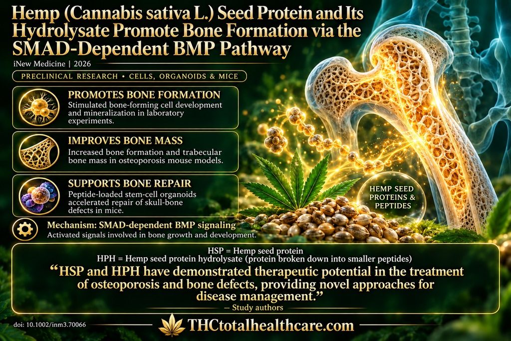
Hemp seed protein and its enzyme-derived peptides promoted bone formation in cell experiments and mouse models of osteoporosis. Researchers found that both preparations stimulated bone-forming cell development and mineral deposition, with the peptide preparation generally producing stronger effects.
These effects involved the SMAD-dependent BMP pathway, a signaling system that helps direct bone growth. Treatment increased bone-formation markers and improved the formation and mass of trabecular bone—the supportive, spongy structure inside bones—in aging and ovariectomy-induced osteoporosis models.
The researchers also incorporated hemp seed peptides into stem-cell-containing organoids, which supported mineralization and accelerated repair after implantation into skull-bone defects in mice. These preclinical findings identify hemp seed proteins and peptides as promising candidates for developing treatments for osteoporosis and bone repair.
“Objective
With the global increase in aging populations, the prevalence of osteoporosis is rising. Current preventive and therapeutic strategies remain limited. The present study aimed to investigate the effects of hemp seed protein (HSP) and its hydrolysis-derived bioactive peptides (HPH) on osteogenic differentiation and to elucidate their therapeutic potential for osteoporosis.
Methods
The osteogenic differentiation of mouse pre-osteoblast MC3T3-E1 cells and human mesenchymal stem cells (hMSCs) was assessed by means of alkaline phosphatase (ALP) staining, alizarin red S (ARS) staining, and RT-qPCR. The screening of signaling pathways was conducted through the implementation of luciferase reporter assays. The in vivo efficacy of the treatment was evaluated in aging and ovariectomy (OVX)-induced osteoporotic mice using calcein labeling and micro-CT. Furthermore, hMSCs encapsulated in HPH-loaded hyaluronic acid methacrylate (HAMA) hydrogel were combined with trabeculae-like biomimetic bone-filling material (TBM) to generate organoids for in vitro culture or calvarial implantation, thereby enabling the evaluation of the in situ osteogenic capacity of HPH.
Results
HSP and HPH enhanced ALP activity, mineralization, and osteogenic gene expression (ALP, RUNX2). Mechanistically, they activated SMAD4 and BMP2/BMP4 expression while suppressing SMAD2, indicating SMAD-dependent BMP pathway involvement. In the mouse models of osteoporosis, the administration of HSP/HPH was found to augment both the mineral apposition rate (MAR) and the trabecular bone mass. In organoid models, HPH was found to maintain cell viability, upregulate ALP and RUNX2 expression, and promote mineralization in vitro. Furthermore, upon calvarial implantation, HPH-loaded organoids were found to accelerate bone defect repair.
Conclusion
These findings indicate that HSP and HPH promote osteogenesis via the SMAD-dependent BMP pathway. The organoid results further demonstrate the in situ osteogenic potential of HPH, supporting its translational applications. HSP and HPH have demonstrated therapeutic potential in the treatment of osteoporosis and bone defects, providing novel approaches for disease management.”