Polyphenol-rich fermented hempseed ethanol extracts improve obesity, oxidative stress, and neural health in high-glucose diet-induced Caenorhabditis elegans

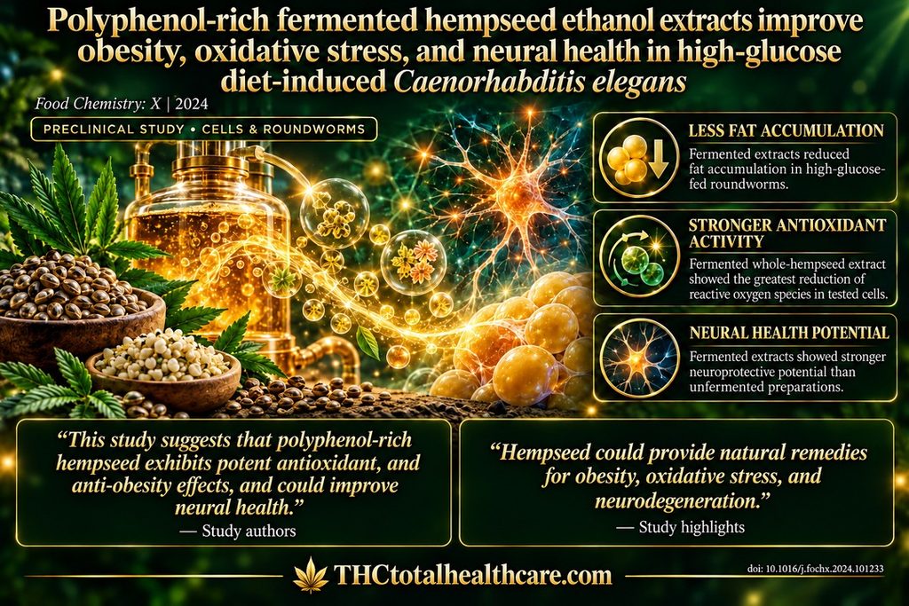
Fermented hempseed extracts reduced fat accumulation and oxidative stress and showed neuroprotective effects in a high-glucose-fed roundworm model. Researchers compared extracts from whole and hulled hemp seeds before and after fermentation, finding that fermented preparations generally produced stronger biological benefits.

Fermented whole-hempseed extract inhibited pancreatic lipase, an enzyme that breaks down dietary fats, by 70.80% and acetylcholinesterase, which breaks down the nerve-signaling chemical acetylcholine, by 78.94% in laboratory assays. It also showed the strongest reduction in reactive oxygen species in tested cells, supporting its antioxidant activity.

The researchers identified diverse seed polyphenols, including flavonoids, phenolic acids, lignanamides, and phenylamides. These preclinical findings highlight fermented hempseed as a promising source of bioactive compounds for research into obesity, oxidative stress, and neural health.

“Whole hempseed (WHS), fermented whole hempseed (FWHS), dehulled hempseed (DHS), and fermented dehulled hempseed (FDHS) ethanol extracts were tested for their toxicity and physiological benefits in relation to their phenolic profiles.

The safety of all samples was confirmed by the absence of toxic effects on HepG2 cells. FWHS exhibited the highest capacity to inhibit lipase activity (70.80%) and acetylcholinesterase (AChE) (78.94%) in vitro. Similarly, in HepG2 cells, FWHS revealed the greatest ability to reduce the accumulation of reactive oxygen species (ROS).

Fermented hempseed demonstrated superior antioxidant, neuroprotective and anti-fat potential, counteracting ageing in high glucose diet-induced C. elegans than unfermented. HPLC and UHPLC-Q-TOF-MS/MS2 phenolic identification revealed the presence of diverse flavonoids, phenolic acids, lignanamides, and phenylamides in hempseed extracts. Among these polyphenols, quercetin, gallic acid, and kaempferol exhibited excellent antioxidant potential, whereas N-trans-feruloyl tyramine displayed the highest anti-lipase potential.

This study suggests that polyphenol-rich hempseed exhibits potent antioxidant, and anti-obesity effects, and could improve neural health.”

https://pubmed.ncbi.nlm.nih.gov/38426074

“Fermented hempseeds demonstrated superior anti-obesity and antioxidant capabilities.”

“Hempseed could provide natural remedies for obesity, oxidative stress, and neurodegeneration.”

https://www.sciencedirect.com/science/article/pii/S2590157524001202?via%3Dihub
Acute Ultra Low Dose THC Attenuates Anxiety-Like Behavior in a Mouse Model for Major-Depressive Disorder

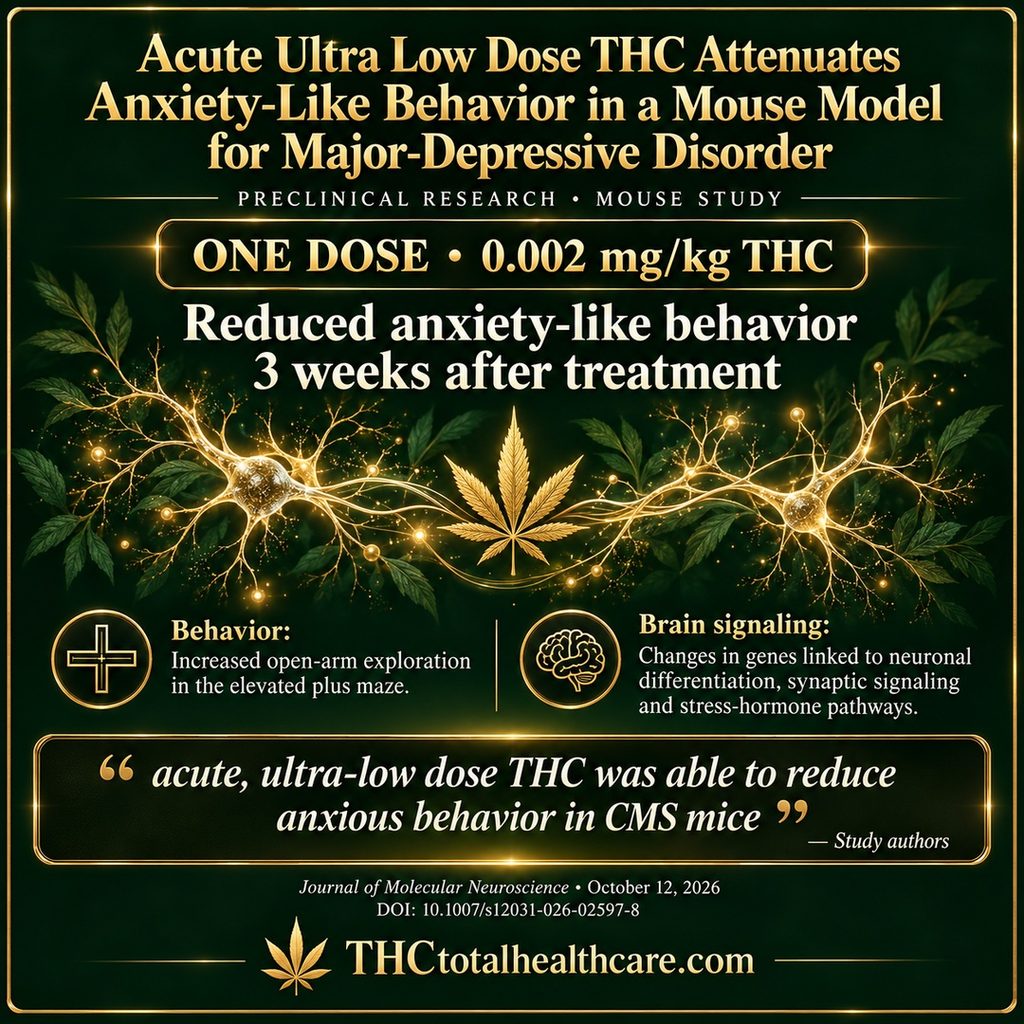
A single ultra-low dose of THC reduced anxiety-like behavior in mice exposed to chronic mild stress, a model used to study major depressive disorder. Researchers administered 0.002 mg/kg of THC and observed behavioral benefits three weeks later, after THC had cleared from the animals’ systems.

THC-treated stressed mice explored the open arms of an elevated plus maze more frequently than untreated stressed mice, indicating reduced anxiety-like behavior. A separate open-field test showed a trend toward improvement. Brain analysis also identified changes in gene expression associated with neuronal differentiation, communication between nerve cells, and stress-hormone signaling.

These findings support further investigation of ultra-low-dose THC for anxiety associated with depression. Changes involving Elmod1 and other stress-related genes offer clues to how a single treatment could produce lasting effects in this mouse model.

“Major depressive disorder (MDD) is a prevalent psychiatric disorder, and is a leading cause of disability worldwide. Current treatments for MDD entail many side effects, and include a lag until the onset of therapeutic effects. The current hypotheses as for the etiology of MDD revolve around the monoamine hypothesis and the plasticity and neurodegeneration hypothesis.

According to the latter, employing a neuroprotective treatment may help ameliorate depressive symptoms. Δ9-Tetrahydrocannabinol (THC) is the primary psychoactive component of the cannabis sativa plant. THC interacts with the endocannabinoid system, which was shown to be involved in the regulation of emotional states (e.g. anxiety) and stress through modulation of synaptic plasticity.

Here, we show the potential of acute, ultra-low dose (0.002 mg/kg) THC in ameliorating anxious behavior in a chronic mild stress mouse model for MDD. We utilize next generation sequencing techniques to show the treatment’s effect on neuronal differentiation and synaptic plasticity-related mRNAs, and specify Elmo domain containing 1 (Elmod1) as a molecular mediator of the observed anxiolytic effects. Moreover, we show the involvement of glucocorticoids-related mRNAs in the observed anxiolytic effect of acute, ultra-low dose THC.”

https://pubmed.ncbi.nlm.nih.gov/42864498

“Acute, Ultra-Low Dose THC Reduces Anxiety in CMS Mice”

“While further studies should be made to deeply understand the molecular mechanism underlying the effects of acute, ultra-low dose THC, with emphasis on the glucocorticoid pathway and cytoskeleton stability, our results show the potential of acute, ultra-low dose THC treatment in ameliorating anxious behavior through distinct mechanisms at the mRNA level. The use of an acute, ultra-low dose holds substantial translational value for clinical adoption, as compliance may be higher when treatment is given only once.

Ultimately, this work shows that an extremely low dose of THC is sufficient to produce behavioral effects without requiring the chronic, high-dose regimens typically needed to achieve similar outcomes.”

https://link.springer.com/article/10.1007/s12031-026-02597-8

Need this THC study explained? thcXplained.com

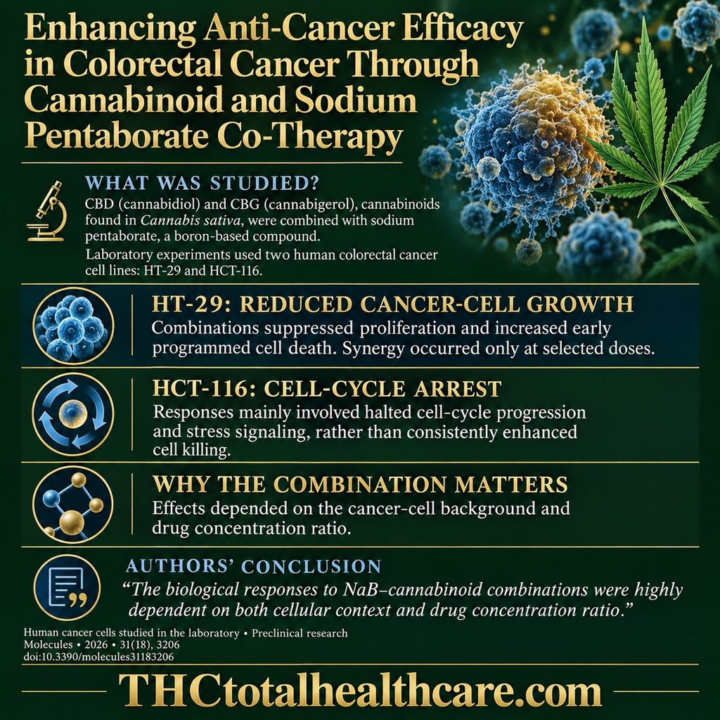
Enhancing Anti-Cancer Efficacy in Colorectal Cancer Through Cannabinoid and Sodium Pentaborate Co-Therapy

Cannabidiol (CBD) and cannabigerol (CBG), naturally occurring cannabinoids found in the Cannabis sativa plant, enhanced growth-suppressing effects at selected doses when combined with sodium pentaborate, a boron-based compound, in laboratory-grown human colorectal cancer cells. Researchers tested two cell lines, HT-29 and HCT-116, and found that responses depended strongly on the cancer cells’ characteristics and the combination used.

In HT-29 cells, the combinations reduced cell proliferation and increased early apoptosis—programmed cell death—with synergistic effects occurring only at certain dose combinations. HCT-116 cells responded mainly by arresting the cell cycle and activating stress-response pathways, rather than showing consistently enhanced cell killing.

The treatments also altered genes involved in oxidative balance and iron metabolism, processes associated with ferroptosis, an iron-dependent form of cell death. These findings support further investigation of boron–cannabinoid combinations tailored to tumor biology, with validation in three-dimensional tumor models and animal studies identified as the next steps.

“Background: Colorectal cancer (CRC) is characterized by pronounced genetic and phenotypic heterogeneity, which substantially influences therapeutic response and limits the efficacy of uniform treatment strategies. Cannabinoid-derived phytochemicals and boron-based compounds have independently been reported to modulate cancer cell proliferation, survival, and redox balance. However, the extent to which these agents interact at the cellular level and whether such interactions are dependent on tumor-specific molecular contexts remains poorly defined.

Methods: Sodium pentaborate (NaB) was combined with non-cytotoxic concentrations of cannabidiol (CBD) or cannabigerol (CBG) and evaluated in HCT-116 and HT-29 colorectal cancer cell lines. Cell viability and drug interactions were assessed by MTS and combination index analyses. Apoptotic responses were examined by Annexin V/PI staining, caspase-3/7 activity assays, and transcriptional profiling of apoptosis-related genes. Cell cycle dynamics and proliferation-associated markers were analyzed by flow cytometry and quantitative PCR. In parallel, ferroptosis-associated gene expression patterns were investigated to evaluate alterations in redox and iron metabolism pathways.

Results: NaB-cannabinoid combinations produced divergent biological outcomes depending on cellular background. HT-29 cells exhibited dose-dependent antiproliferative responses to NaB + CBD and NaB + CBG, with synergistic interactions observed only at selected dose combinations accompanied by increased early apoptosis. In contrast, HCT-116 cells primarily responded with cell cycle arrest and transcriptional stress signaling rather than enhanced cytotoxicity. Modulation of ferroptosis-related gene expression further indicated differential redox adaptation between the two cell models.

Conclusions: NaB-cannabinoid combinations elicit distinct biological outcomes in colorectal cancer cells that are strongly determined by cellular context. While HT-29 cells are selectively sensitized to combination dose level, HCT-116 cells predominantly respond through cell cycle arrest and adaptive stress-response pathway activation. These findings emphasize the necessity of context-aware combination strategies and provide a mechanistic framework for the further development of boron-cannabinoid-based therapeutic approaches in colorectal cancer.”

https://pubmed.ncbi.nlm.nih.gov/42796493

“In conclusion, our findings indicate that co-treatment with NaB and phytocannabinoids provides a cell line-specific therapeutic strategy in colorectal cancer.”

https://www.mdpi.com/1420-3049/31/18/3206

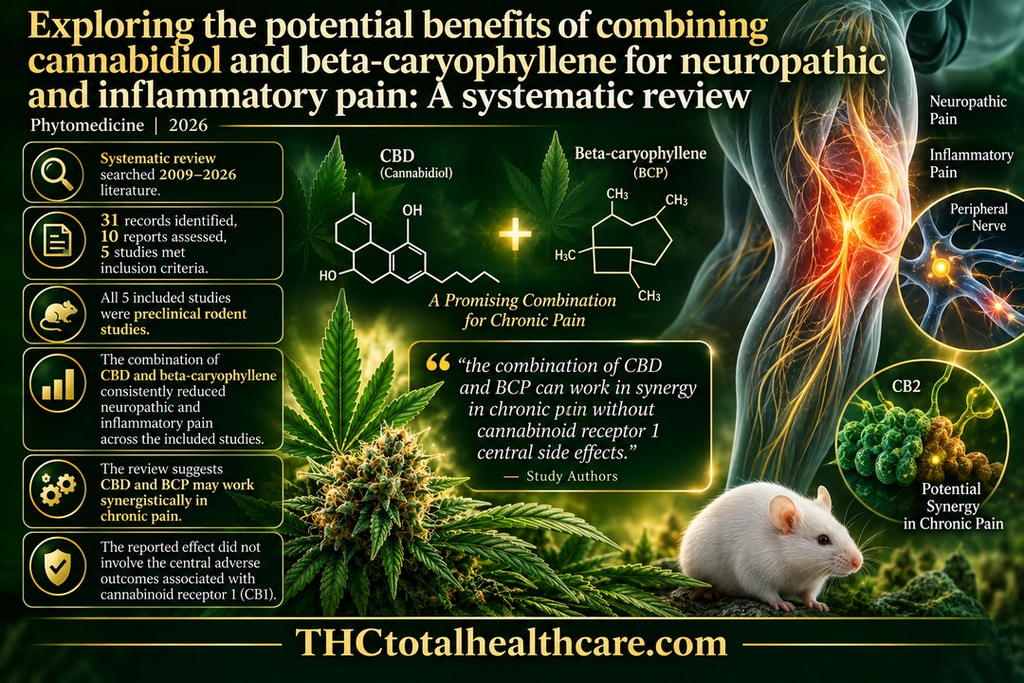
Exploring the potential benefits of combining cannabidiol and beta-caryophyllene for neuropathic and inflammatory pain: A systematic review

A new systematic review found that combining cannabidiol (CBD) with beta-caryophyllene (BCP) consistently reduced neuropathic and inflammatory pain in preclinical studies. The review searched research published from 2009 through 2026 and identified five studies that met its inclusion criteria, all using rodent pain models.

Across those studies, the CBD–BCP combination reduced both neuropathic and inflammatory pain, with the authors describing evidence of a possible synergistic analgesic effect. Importantly, the reported pain relief was not accompanied by the central adverse effects associated with cannabinoid receptor 1 (CB1) activation.

The authors concluded that CBD and beta-caryophyllene may work together synergistically in chronic pain and said the findings provide a rationale for future preclinical and clinical studies. Because the evidence so far is entirely preclinical, the next step is determining whether the same combination can produce meaningful pain relief in people.

“Purpose: In this review, we examined the analgesic effect of the combination of cannabidiol (CBD) and beta-caryophyllene (BCP) in different pain conditions.

Methods: A systematic review was conducted in accordance with the Preferred Reporting Items for Systematic Reviews and Meta-Analyses guidelines. Literature searches were performed across major academic databases (Google Scholar, Web of Science, and PubMed) from 2009 to 2026 to identify studies investigating the analgesic effect of the combination of CBD and BCP.

Results: The search identified 31 records. Ten reports were screened and assessed for eligibility, and five studies met the inclusion and exclusion criteria. Findings consistently demonstrate that the combination of CBD and BCP reduces neuropathic and inflammatory pain in rodent models. This effect does not result in adverse outcomes associated with cannabinoid receptor 1 (CB1). This review discusses potential mechanisms by which this combination produces synergistic analgesic effects.

Conclusion: The literature discussed in this review suggests that the combination of CBD and BCP can work in synergy in chronic pain without cannabinoid receptor 1 central side effects. These preclinical findings provide the rationale for future preclinical and clinical studies.”

https://pubmed.ncbi.nlm.nih.gov/42753475

“In summary, these data suggest that combining CBD and BCP may be especially effective in reducing neuropathic pain caused by spinal cord injury. Additionally, this combination resulted in minimal cannabinoid-like side effects.”

https://www.sciencedirect.com/science/article/pii/S0944711326010287?via%3Dihub

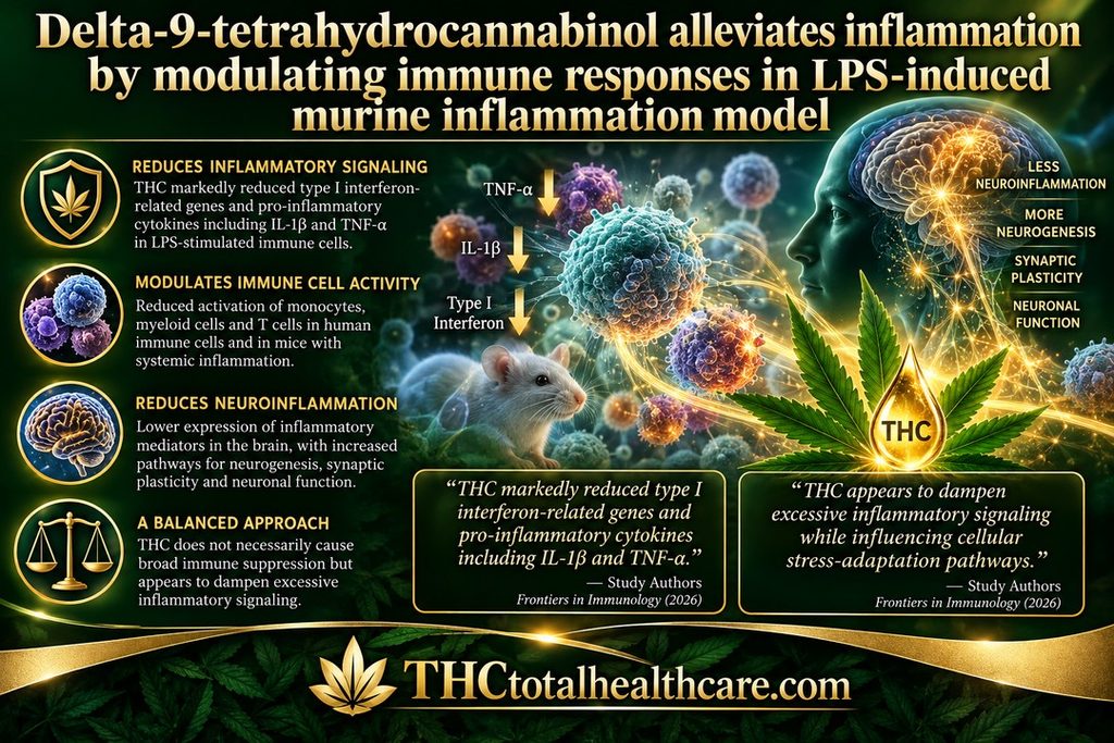
Delta-9-tetrahydrocannabinol alleviates inflammation by modulating immune responses in LPS-induced murine inflammation model

Delta-9-THC reduced inflammatory immune signaling across human immune cells and a mouse model of systemic inflammation. In LPS-stimulated cells, THC markedly suppressed type I interferon-related genes and pro-inflammatory cytokines including IL-1β and TNF-α, while also reducing immune-cell activation markers.

The effects extended beyond isolated cells. In mice exposed to systemic inflammation, THC reduced activation of splenic myeloid cells and T cells. Brain analysis also showed reduced neuroinflammatory signaling, including lower expression of inflammatory mediators, while pathways associated with neurogenesis, synaptic plasticity and neuronal function were enriched.

Importantly, the researchers describe THC’s effects as modulation rather than indiscriminate immune suppression, concluding that THC appears to dampen excessive inflammatory signaling while influencing cellular stress-adaptation pathways. They say these findings support further investigation of cannabinoid signaling in chronic inflammatory, neuroimmune and neurodegenerative disorders.

“Introduction: Δ9-Tetrahydrocannabinol (THC), the primary psychoactive component of cannabis, has been reported to modulate immune responses; however, its effects on complex innate immune signaling pathways and inflammation remain incompletely understood. Here, we investigated the immunomodulatory effects of THC on lipopolysaccharide (LPS)-induced inflammation across cellular and in vivo models.

Methods: We integrated transcriptomic, cellular, and in vivo approaches to characterize the effects of THC on innate immune activation. Human THP-1 monocytic cells were treated with THC in the presence or absence of LPS, followed by transcriptomic and gene expression analyses to evaluate inflammatory, type I interferon, unfolded protein response (UPR), and autophagy pathways. Findings were validated in primary human monocytes by assessing immune activation markers. The effects of chronic THC exposure were further evaluated in a murine model of systemic inflammation by examining splenic myeloid and T-cell activation. RNA sequencing of brain tissue was performed to assess the effects of THC on neuroinflammatory and neuronal pathways.

Results: THC induced a stress-adaptive transcriptional program in THP-1 cells, characterized by upregulation of genes associated with the UPR and autophagy pathways. In contrast, THC markedly suppressed LPS-induced type I interferon-stimulated genes (ISGs) and pro-inflammatory cytokines, including IL-1β and TNF-α. In primary human monocytes, THC significantly reduced the expression of activation markers CD80, CD83, and CD209. Consistent with these findings, chronic THC exposure attenuated the activation of splenic myeloid and T cells in mice subjected to systemic inflammation. Brain transcriptomic analysis further demonstrated reduced expression of neuroinflammatory pathways following THC exposure, accompanied by enrichment of pathways associated with neurogenesis and synaptic plasticity.

Discussion: Our findings demonstrate that THC exerts broad immunomodulatory effects characterized by suppression of inflammatory and type I interferon responses while promoting cellular stress-adaptation pathways. These effects were observed across human monocytic cells, primary monocytes, and peripheral immune populations in vivo, and were accompanied by reduced neuroinflammatory signaling and increased neuronal plasticity-associated pathways in the brain. Together, these findings provide mechanistic insight into the immunomodulatory actions of THC and support further investigation of cannabinoid signaling as a potential regulator of chronic inflammatory and neuroimmune disorders.”

https://pubmed.ncbi.nlm.nih.gov/42818626

“Chronic THC exposure reduces LPS-induced immune activation in vivo.”

https://www.frontiersin.org/journals/immunology/articles/10.3389/fimmu.2026.1856131/full

Need this THC study explained? thcXplained.com

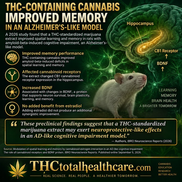
Modulation of spatial learning and memory by cannabinoid-estrogen interaction in an AD-like cognitive impairment: The role of cannabinoid receptors and BDNF protein

A 2026 study found that a THC-standardized marijuana extract improved spatial learning and memory in an Alzheimer’s-like model of cognitive impairment. Researchers tested the extract in rats exposed to amyloid-beta, a protein closely associated with Alzheimer’s disease, and observed improvements in memory performance.

The cognitive improvements were accompanied by changes in cannabinoid receptor signaling and in BDNF, or brain-derived neurotrophic factor, a protein that supports neuron survival, brain plasticity, learning, and memory. The findings suggest that the brain’s cannabinoid system and BDNF may be involved in the neuroprotective-like effects observed with the THC-standardized extract.

The researchers also examined the interaction with estrogen and found that adding estradiol did not produce an additional synergistic improvement. As the authors concluded, “These preclinical findings suggest that a THC-standardized marijuana extract may exert neuroprotective-like effects in an AD-like cognitive impairment model.”

“Alzheimer’s disease (AD) is an increasingly prevalent neurodegenerative disorder worldwide, with women showing a higher risk of developing the disease. The decline in steroid hormones after menopause may contribute to the increased susceptibility of women to neurodegenerative conditions.

In parallel, cannabis-derived compounds have been reported to alleviate certain symptoms associated with neurological disorders. The present study was designed to investigate the effects of marijuana extract on cognitive impairment and hippocampal molecular markers in an ovariectomized AD-like rat model, and to evaluate whether co-administration with estradiol modifies these effects.

The marijuana extract used in this study was characterized by HPLC and was found to contain 8.5% Δ9-THC. AD-like cognitive impairment pathology was induced by intra-hippocampal administration of Aβ25-35 in OVX rats. Animals were treated with marijuana extract (60 mg/kg/day, corresponding to approximately 5.1 mg/kg/day Δ9-THC) either alone or in combination with 17β-estradiol (1 mg/kg every 4 days) for 28 days. Cognitive performance was evaluated using the Morris water maze (MWM). In addition, hippocampal CB1/CB2 receptor expression and BDNF protein levels were measured to examine potential molecular associations.

Our findings showed that chronic administration of the marijuana extract improved Aβ25-35-induced deficits in spatial learning and memory.

Alterations in CB1 receptor expression and BDNF levels accompanied these behavioral effects. Notably, co-treatment with estradiol did not produce a synergistic effect, suggesting a complex interaction between cannabinoid-related and estrogen-related signaling pathways.

These preclinical findings suggest that a THC-standardized marijuana extract may exert neuroprotective-like effects in an AD-like cognitive impairment model. However, because the extract was not fully phytochemically characterized and the mechanistic analyses were correlational, the results should be interpreted cautiously. Further studies are needed to clarify the underlying mechanisms and translational relevance of these findings.”

https://pubmed.ncbi.nlm.nih.gov/42756662

“In conclusion, chronic administration of THC-standardized marijuana extract improved spatial learning and memory performance in the AD-like cognitive impairment model, and these behavioral changes were accompanied by alterations in hippocampal CB1/CB2 gene expression and BDNF protein levels.”

https://www.ibroneuroreports.org/article/S2667-2421(26)00149-1/fulltext

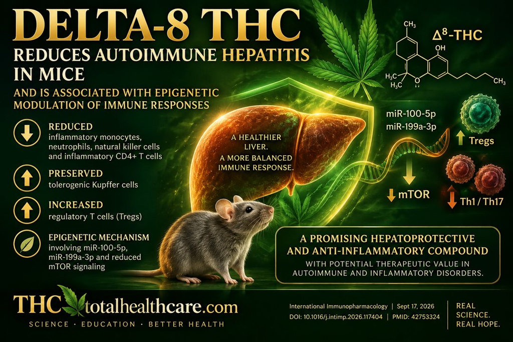
Delta-8-tetrahydrocannabinol ameliorates murine autoimmune hepatitis and is associated with epigenetic modulation of immune responses

A new 2026 study published in International Immunopharmacology found that delta-8-tetrahydrocannabinol (Δ8-THC) significantly reduced autoimmune hepatitis in mice while altering the immune responses responsible for liver inflammation. Researchers found that Δ8-THC reduced inflammatory immune cells and helped preserve immune cells involved in protecting the liver.

The researchers also uncovered a possible mechanism. Δ8-THC increased two microRNAs, miR-100-5p and miR-199a-3p, which suppressed mTOR signaling and shifted CD4+ T-cell development away from inflammatory Th1 and Th17 cells and toward regulatory T cells that help control excessive immune activity.

The authors concluded that Δ8-THC showed both hepatoprotective and anti-inflammatory activity and highlighted its potential therapeutic value for autoimmune and inflammatory disorders. The study adds new evidence that cannabinoids may influence disease not only by reducing inflammation, but by changing the molecular and epigenetic pathways that regulate immune responses.

“Aim of the study: Cannabinoids have gained attention for their ability to modulate immune responses and suppress inflammation. However, the effect of Δ8-THC, a minor cannabinoid found in Cannabis, has not been explored in autoimmune hepatitis. To address knowledge gap, the present study aimed to investigate the hepatoprotective and immunoregulatory properties of Δ8-THC in experimental autoimmune hepatitis and to identify molecular and epigenetic mechanisms underlying its immunoregulatory effects.

Methods: miRNA sequencing and flow cytometric analysis were performed using hepatic immune cells from female C57BL/6 mice treated with ConA (12.5 mg/kg, i.v.) and Δ8-THC (20 mg/kg, i.p.). Differential miRNA expressions were analyzed using the edgeR, and miRNA targets were predicted using Ingenuity Pathway Analysis. Target gene expression was validated by qRT-PCR and Western blot. Functional assays including miRNA mimic/inhibitor transfection and CD4+ T-cell differentiation were performed to validate target gene regulation by miRNAs and its role in CD4+ T-cell differentiation.

Results: Δ8-THC markedly suppressed ConA-induced autoimmune hepatitis by restoring immune homeostasis within the liver through suppression of inflammatory monocytes, neutrophils, and natural killer cells, and preservation of tolerogenic Kupffer cells. In addition, Δ8-THC reshaped the hepatic lymphoid compartment, diminishing inflammatory CD4+ T-cell responses while favoring regulatory T-cell expansion. These effects were associated with the preservation of miR-100-5p and miR-199a-3p expression in the liver, whereas both miRNAs were markedly downregulated following ConA treatment. mTOR, a central regulator of T-cell differentiation, was identified as a target of these miRNAs. Consistent with these findings, Δ8-THC reduced mTOR expression and upregulated hepatoprotective miRNAs in both in vivo and in vitro models. Functional analyses with miRNA overexpression and inhibition approaches verified the direct regulation of mTOR by these miRNAs in T cells and RAW 264.7 cells. In naïve CD4+ T cells, miRNA gain- and loss-of-function experiments demonstrated that miR-100-5p/miR-199a-3p-mediated suppression of mTOR promotes regulatory T-cell differentiation while limiting Th1 and Th17 polarization.

Conclusion: Our results identify Δ8-THC as a promising hepatoprotective and anti-inflammatory compound that ameliorates ConA-induced autoimmune hepatitis by influencing CD4+ T-cell differentiation potentially through the miR-100-5p/miR-199a-3p-mTOR axis, highlighting its potential therapeutic value in autoimmune and inflammatory disorders.”

https://pubmed.ncbi.nlm.nih.gov/42753324

“In conclusion, our findings suggest that Δ8-THC can mitigate ConA-induced hepatitis, potentially through epigenetic regulation involving microRNAs. Specifically, Δ8-THC improved liver function and reduced systemic and intrahepatic inflammation in ConA-exposed mice. These effects were associated with increased expression of miR-100-5p and miR-199a-3p in the liver, suggesting that these miRNAs may regulate mTOR signaling and contribute to the suppression of proinflammatory responses and promotion of anti-inflammatory pathways. Functional studies supported the involvement of these miRNAs in the regulation of mTOR signaling, CD4+ T-cell differentiation, and inflammatory responses.”

https://www.sciencedirect.com/science/article/pii/S1567576926012518?via%3Dihub

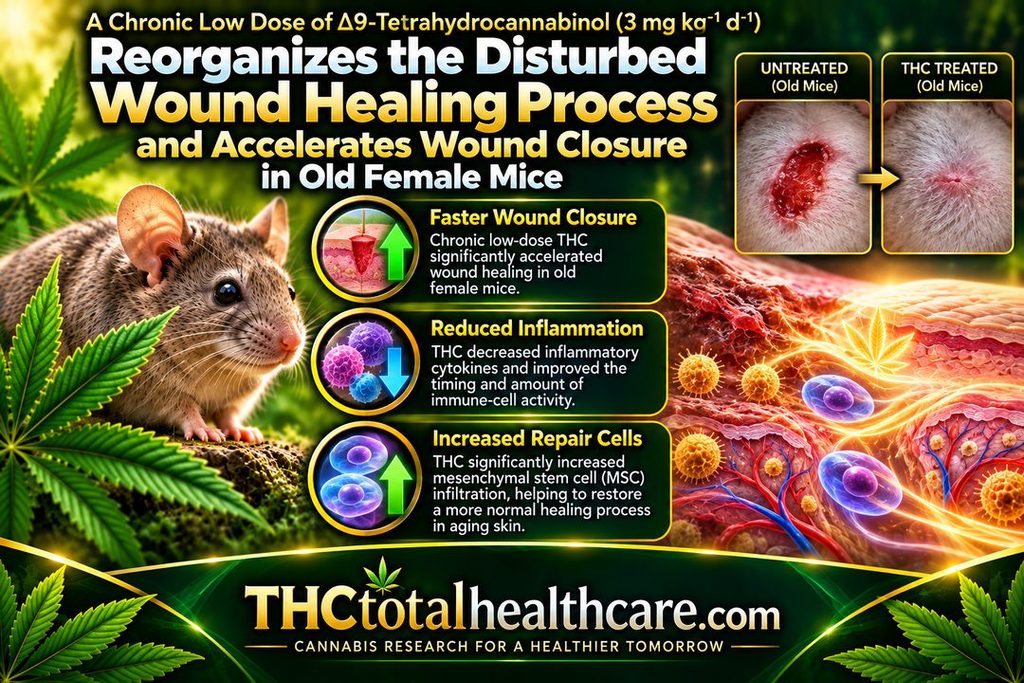
A chronic low dose of Δ9-tetrahydrocannabinol (3 mg / kg / 21 d) reorganizes the disturbed wound healing process and accelerates wound closure in old female mice

A chronic low dose of Δ9-THC accelerated wound closure in old female mice and helped reorganize an age-disrupted healing process. The biggest improvement occurred during the inflammatory phase, when aged wounds normally heal more slowly and remain inflamed longer.

THC changed the timing and amount of immune-cell activity in the wound, reduced inflammatory cytokines, and significantly increased infiltration of mesenchymal stem cells (MSCs) involved in tissue repair. The treatment did not increase growth-factor release, suggesting the benefit came mainly from better coordination of inflammation and cell migration.

The researchers concluded that THC improved wound healing by restoring a more normal sequence of immune-cell infiltration and cytokine release. The findings suggest that stimulating the aging endocannabinoid system may help support skin regeneration and tissue repair in older organisms.

“Wound healing in old mice is characterized by disturbed tissue homeostasis, manifested by delayed immune cell infiltration and reduced growth factor secretion, leading to a delayed onset and prolonged duration of the inflammatory phase.

The endocannabinoid system (ECS) is an important regulator of tissue homeostasis and cell migration and is also considered to be subject to aging processes, which may contribute to observable aging phenomena. Therefore, stimulating the aged ECS could represent a therapeutic option to support tissue regeneration in aging.

Female old mice received a low-dose of medical THC daily for 3 weeks, before four excisional full skin wounds were created. At day 1, 3 and 7 post-surgery, the wound closure rate was analyzed and wound samples were examined immunohistochemically for the numbers of granulocytes, M1-macrophages and mesenchymal stem cells (MSCs). The concentrations of inflammatory cytokines and regenerative growth factors were determined by ELISA.

Administration of THC improved the wound healing rate of old mice between day 1 and 7, which was associated with an altered timing and quantity of infiltrating immune cells and decreased levels of inflammatory cytokines in wound tissue on days 1 and 3 post-injury.

THC treatment significantly increased MSC infiltration but had no effect on the growth factor release.

The present study confirmed the anti-inflammatory activity of THC in vivo.

The THC-treatment improved wound healing in old mice by coordinating the temporal sequence of immune cell infiltration and cytokine release. Thus, restoration of ECS signaling could be an effective strategy to support age-related skin regeneration.”

https://pubmed.ncbi.nlm.nih.gov/40653209/

“A chronic low dose of THC accelerates the wound closure rate during the inflammatory phase in old mice.”

https://www.sciencedirect.com/science/article/pii/S0531556525001615?via%3Dihub

Meed this THC study explained? thcXplained.com

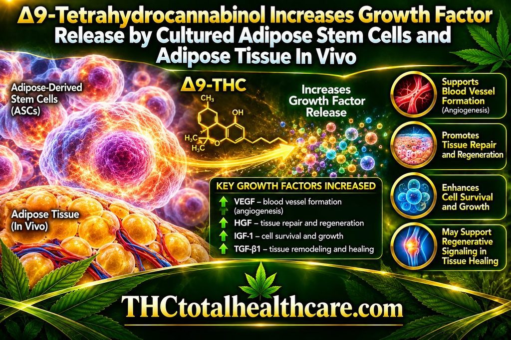
∆9-Tetrahydrocannabinol Increases Growth Factor Release by Cultured Adipose Stem Cells and Adipose Tissue in vivo

Δ9-THC increased the release of growth factors from cultured adipose-derived stem cells and from adipose tissue in vivo. These growth factors are involved in processes such as cell survival, tissue repair, blood-vessel formation, and regeneration.

The researchers found that THC changed the secretory activity of adipose stem cells rather than simply affecting their growth. This suggests that THC may influence how these cells communicate with surrounding tissue and support repair through the factors they release.

The findings point to a potential role for THC in regenerative signaling and tissue-healing biology, particularly through adipose-derived stem cells and the local tissue environment. The study adds another dimension to cannabinoid research by showing that THC can affect not just mature cells, but also the reparative signals produced by stem-cell populations.

“Background: Because of its biocompatibility and its soft and dynamic nature, the grafting of adipose tissue is regarded an ideal technique for soft-tissue repair. The adipose stem cells (ASCs) contribute significantly to the regenerative potential of adipose tissue, because they can differentiate into adipocytes and release growth factors for tissue repair and neovascularization to facilitate tissue survival. The present study tested the effect of administering a chronic low dose of ∆9-tetrahydrocannabinol (THC) on these regenerative properties, in vitro and in vivo.

Methods: Human ASCs were exposed to increasing concentrations of THC. Resazurin conversion was applied to investigate the effect on metabolic activity, cell number was assessed by crystal violet staining, tri-linear differentiation was evaluated by specific colorimetric approaches, and the release of growth factors was analyzed by ELISA. Two groups of mice were treated daily either with a low dose of THC (3 mg/kg) or a vehicle solution. After 3 weeks, adipose tissue was obtained from excised fat deposits, homogenized and tested for growth factor contents.

Results: THC decreased ASC proliferation but increased metabolic activity as well as adipogenic and chondrogenic differentiation. A low concentration of THC (1 µM) enhanced the growth factor release by ASCs. The concentration of these cytokines was also increased in adipose tissue of mice treated with THC.

Conlusion: Our results indicate that chronic activation of the endocannabinoid system promoted differentiation and growth factor release of ASCs, which could be of specific value for enhancing the regenerative potential of adipose tissue.”

https://pubmed.ncbi.nlm.nih.gov/39825991/

“Our findings may offer a new approach for improved application of adipose tissue in regenerative medicine.”

https://link.springer.com/article/10.1007/s13770-024-00692-8

Need this THC study explained? thcXplained.com

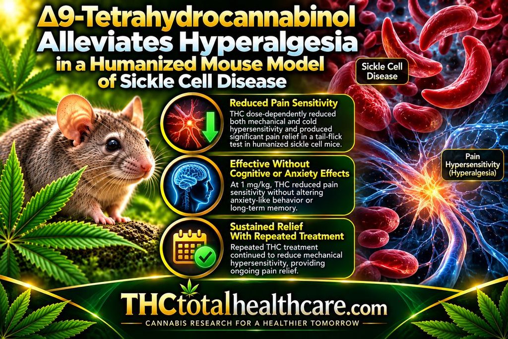
Δ 9 -Tetrahydrocannabinol alleviates hyperalgesia in a humanized mouse model of sickle cell disease

Δ9-THC reduced pain hypersensitivity in a humanized mouse model of sickle cell disease. In mice expressing more than 99% human sickle hemoglobin, acute THC treatment dose-dependently reduced both mechanical and cold hypersensitivity and also produced substantial antinociceptive effects in a tail-flick pain test.

At 1 mg/kg, THC reduced pain sensitivity without altering anxiety-like behavior or long-term memory. With repeated treatment, THC continued to provide sustained relief from mechanical hypersensitivity, although tolerance developed to its effects on cold hypersensitivity.

The researchers concluded that THC may represent a potential option for managing chronic pain associated with sickle cell disease. These findings are especially notable because sickle cell pain is often severe and difficult to control, while current treatment frequently depends on opioid analgesics.

“People with sickle cell disease (SCD) often experience chronic pain as well as unpredictable episodes of acute pain, which significantly affect their quality of life and life expectancy. Current treatment strategies for SCD-associated pain primarily rely on opioid analgesics, which have limited efficacy and cause serious adverse effects.

Cannabis has emerged as a potential alternative, yet its efficacy remains uncertain. In this study, we investigated the antinociceptive effects of Δ9-tetrahydrocannabinol (THC), cannabis’ intoxicating constituent, in male HbSS mice, which express >99% human sickle hemoglobin, and male HbAA mice, which express normal human hemoglobin A, as a control.

Acute THC administration (0.1-3 mg-kg-1, intraperitoneal, i.p.) dose-dependently reduced mechanical and cold hypersensitivity in HbSS, but not HbAA mice. In the tail-flick assay, THC (1 and 3 mg-kg-1, i.p.) produced substantial antinociceptive effects in HbSS mice. By contrast, THC (1 mg-kg-1, i.p.) did not alter anxiety-like behavior (elevated plus maze) or long-term memory (24-h novel object recognition). Subchronic THC treatment (1 and 3 mg-kg-1, i.p.) provided sustained relief of mechanical hypersensitivity but led to tolerance in cold hypersensitivity in HbSS mice.

Together, the findings identify THC as a possible therapeutic option for the management of chronic pain in SCD. Further research is warranted to elucidate its mechanism of action and possible interaction with other cannabis constituents. 

Significance Statement The study explores THC’s efficacy in alleviating pain in sickle cell disease (SCD) using a humanized mouse model. Findings indicate that acute THC administration reduces mechanical and cold hypersensitivity in SCD mice without impacting emotional and cognitive dysfunction. Subchronic THC treatment offers sustained relief of mechanical hypersensitivity but leads to cold hypersensitivity tolerance. These results offer insights into THC’s potential as an alternative pain management option in SCD, highlighting both its benefits and limitations.”

https://pubmed.ncbi.nlm.nih.gov/38955494/

“The results indicate that acute THC administration alleviates mechanical and cold hypersensitivity in HbSS mice in a dose-dependent manner without affecting emotional and cognitive dysfunction also present in these mice.”

https://www.sciencedirect.com/science/article/abs/pii/S0022356524178147

Need this THC study explained? thcXplained.com