Brain cannabinoid CB1 receptor signaling modulates reward responses and inhibitory control in humans

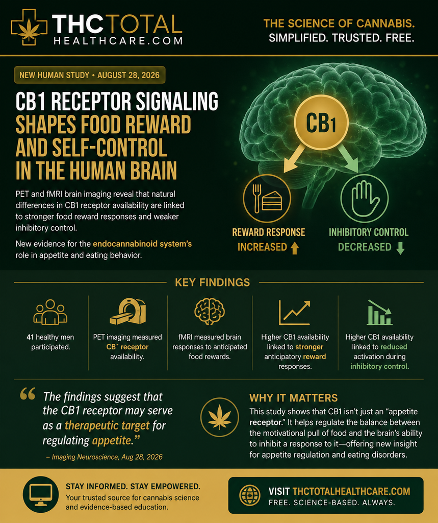
Researchers using PET and fMRI brain imaging found that natural differences in cannabinoid CB1 receptor availability are associated with how strongly the brain responds to anticipated food rewards and how it engages inhibitory-control systems. The findings provide new human evidence connecting the endocannabinoid system with appetite-related behavior and suggest CB1 signaling as a potential therapeutic target for eating disorders and appetite regulation.

“The central endocannabinoid system, particularly the in vivo cannabinoid type 1 (CB1) receptor signaling, presents a promising target for treating eating disorders. However, its precise role in appetite control remains unclear.

This study aimed to determine how CB1 receptor signaling contributes to key aspects of appetite regulation, specifically anticipatory food reward responses and inhibitory control.

Forty-one healthy male participants underwent [18F]FMPEP-d2 positron emission tomography (PET) to quantify CB1 receptor availability. Functional magnetic resonance imaging (fMRI) was used to assess anticipatory neural responses to food cues, while inhibitory control was measured using a go/nogo task.

Data show that individuals with higher CB1 receptor availability exhibited stronger anticipatory reward-related neural responses and reduced activation during inhibitory control. Therefore, CB1 receptor signaling plays a distinct role in modulating reward and inhibitory processes related to feeding behavior.

The findings suggest that the CB1 receptor may serve as a therapeutic target for regulating appetite.”

https://pubmed.ncbi.nlm.nih.gov/42662580

“Brain cannabinoid CB1 receptor availability modulates reward responses and inhibitory control, key neural mechanisms involved in appetite regulation. The findings suggest that individuals with elevated CB1 receptor availability may require increased engagement of inhibitory control mechanisms to counteract reward-driven responses triggered by external cues, as opposed to enhanced internal satiety signals.

These insights have potential implications for developing therapeutic strategies targeting the endogenous cannabinoid system to address obesity epidemic.”
https://direct.mit.edu/imag/article/doi/10.1162/IMAG.a.1344/138067/Brain-cannabinoid-CB1-receptor-signaling-modulates

Multiple binding modes underlie Cannabis sativa cannabinoids recognition by peroxisome proliferator-activated receptor gamma

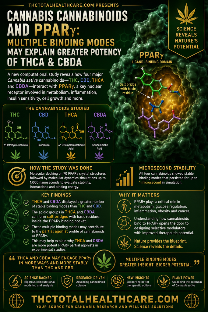
Cannabinoids interact with more than the classical cannabinoid receptors CB1 and CB2. One important target is PPARγ, a nuclear receptor involved in metabolism, inflammation, insulin sensitivity and several disease-related pathways.

In this study, researchers examined how multiple Cannabis sativa cannabinoids bind to and activate PPARγ. The results showed that different cannabinoids can engage the receptor through multiple binding modes, with acidic cannabinoids such as THCA and CBDA showing particularly strong activity.

The findings help clarify another molecular pathway through which cannabis compounds may influence biological processes relevant to metabolic, inflammatory and other disorders.

Introduction: Peroxisome proliferator-activated receptor gamma (PPARγ) is a ligand-activated nuclear receptor with broad therapeutic relevance across various pathologies, including type 2 diabetes, obesity, cancer, and inflammatory disorders. Cannabinoids are a class of terpene-phenolic compounds from Cannabis sativa L. that have been shown to act as partial agonists of PPARγ. Among them, the acidic forms Δ9-tetrahydrocannabinolic acid (THCA) and cannabidiolic acid (CBDA) display higher potency than their decarboxylated counterp arts Δ9-tetrahydrocannabinol (THC) and cannabidiol (CBD). Despite experimental evidence supporting direct PPARγ-cannabinoid interaction, the molecular determinants governing ligand recognition within the binding pocket have not yet been comprehensively investigated.

Methods: A combination of molecular docking and molecular dynamics simulations was employed to characterize the binding modes of THC, CBD, THCA, and CBDA within the PPARγ ligand-binding domain. Docking calculations were performed on a curated set of 70 PPARγ crystal structures co-crystallized with structurally diverse ligands, exploiting thus the conformational variability of the binding pocket. The best-ranked solutions were subjected to 500 ns MD simulations and evaluated on the basis of ligand stability, persistence of polar and aromatic-aromatic interactions with the receptor, and energetic contributions estimated by MM/GBSA. Three candidate binding modes per ligand were selected and their trajectories extended to 1,000 ns.

Results: All four cannabinoids yielded at least one stable binding mode at the microsecond timescale. The cannabinoids THCA and CBDA displayed a greater number of stable binding modes than THC and CBD, a result consistent with the higher potency previously reported for these compounds in experimental studies. This behavior may be attributable to the formation of salt bridges with basic residues in the binding pocket.

Conclusion: Our findings provide a structural framework for understanding cannabinoid recognition by PPARγ. The ability of these compounds to adopt multiple binding modes may contribute to their partial agonist profile, opening new avenues for the rational design of selective PPARγ modulators with improved therapeutic properties.”

https://pubmed.ncbi.nlm.nih.gov/42639023

“This work provides a foundation for future studies addressing the molecular basis of cannabinoid action on PPARγ.

Moreover, these findings could serve as a starting point to explore the allosteric mechanism by which these small lipophilic molecules influence receptor structure and dynamics.

Finally, the strategy described here may also be applied to determine the binding modes of these natural compounds in other members of the nuclear receptor family, including PPARα and PPARδ, as well as to investigate the binding modes of minor cannabinoids, such as cannabichromene and cannabigerol.”

https://www.frontiersin.org/journals/bioinformatics/articles/10.3389/fbinf.2026.1893303/full


The beneficial properties of CBDA in diet-induced neuroinflammation

Cannabidiolic acid (CBDA), the natural acidic precursor of CBD, is drawing increasing attention for its own biological effects.

In this study, researchers examined CBDA in a model of diet-induced neuroinflammation and found evidence that it may help reduce inflammatory changes in the brain associated with an unhealthy diet.

The findings add to growing research showing that acidic cannabinoids such as CBDA may have therapeutic properties distinct from their better-known neutral forms.

“Cannabidiolic acid (CBDA) is a phytocannabinoid found in the Cannabis plant. Understanding the effects of CBDA is essential to uncover its full potential and possible health benefits.

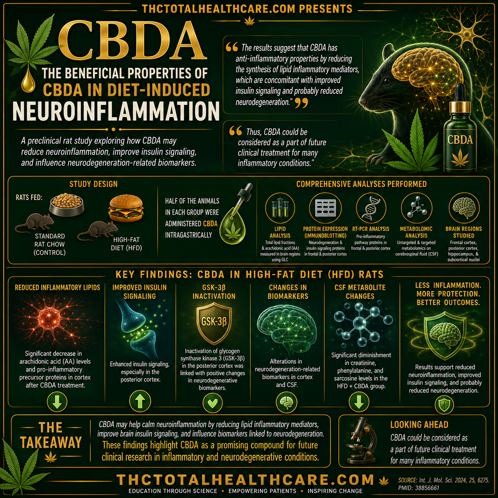
The study was conducted on rats receiving standard rat chow (control) and a high-fat diet (HFD).

Half of the animals in each group were administered CBDA intragastrically. The total lipid fractions and arachidonic acid (AA) contents were measured in the frontal and posterior cortex, hippocampus, and subcortical nuclei using gas-liquid chromatography. The expression of proteins involved in neurodegenerative diseases and insulin signaling pathway proteins in the frontal and posterior cortex was measured using Immunoblotting. RT-PCR was used to assess the expression of pro-inflammatory pathway proteins in the same regions. Additionally, untargeted and targeted metabolomic analyses were performed on cerebrospinal fluid (CSF).

The results showed that a decrease in arachidonic acid levels and pro-inflammatory precursor proteins after CBDA treatment in high-fat-fed rats was simultaneous with improved insulin signaling, particularly in the posterior cortex.

Inactivation of glycogen synthase kinase 3 (GSK-3β) in this region was concomitant with changes in neurodegenerative biomarkers in the cortex and CSF. Metabolomic studies revealed a significant diminishment in creatinine, phenylalanine, and sarcosine levels in the HFD+CBDA group, suggesting it plays an important role in neurological disorders.

The results suggest that CBDA has anti-inflammatory properties by reducing the synthesis of lipid inflammatory mediators, which are concomitant with improved insulin signaling and probably reduced neurodegeneration.

Thus, CBDA could be considered as a part of future clinical treatment for many inflammatory conditions.”

https://pubmed.ncbi.nlm.nih.gov/42627619

“Our study demonstrated a preliminary analysis of the impact of CBDA on the inflammatory profile of the brain under conditions of excess calories from fat. The results suggest an anti-inflammatory role for this cannabinoid, particularly in inhibiting the synthesis of lipid inflammatory mediators.”

https://link.springer.com/article/10.1007/s10787-026-02358-4

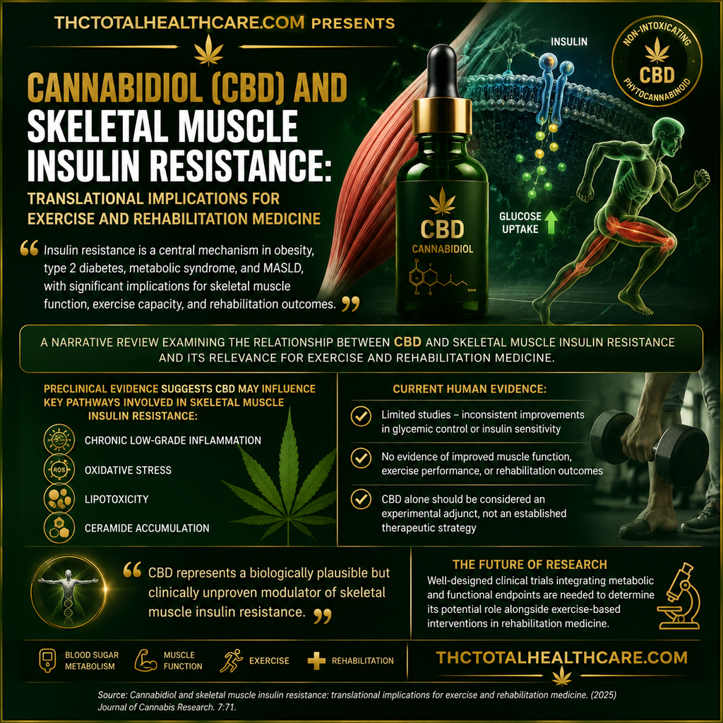
Cannabidiol and skeletal muscle insulin resistance: translational implications for exercise and rehabilitation medicine

Cannabidiol (CBD) is being studied for effects that extend beyond the nervous system, including its potential influence on metabolism and skeletal muscle function.

This review examines how CBD may affect insulin resistance in skeletal muscle, with particular attention to inflammation, oxidative stress, mitochondrial function and glucose metabolism. It also considers how these effects could relate to exercise and rehabilitation medicine.

The findings highlight a potential role for CBD in metabolic health and muscle recovery that warrants further investigation.

“Insulin Resistance (IR) is a central pathophysiological mechanism underlying obesity, type 2 diabetes mellitus, metabolic syndrome, and metabolic dysfunction-associated steatotic liver disease, with significant implications for skeletal muscle function, exercise capacity, and rehabilitation outcomes. Because skeletal muscle represents the primary site of insulin-stimulated glucose disposal, muscle insulin resistance is a key determinant of both metabolic health and physical performance.

Cannabidiol (CBD), a non-intoxicating phytocannabinoid derived from Cannabis sativa, has attracted increasing scientific interest due to its anti-inflammatory, antioxidant, and pleiotropic signaling properties.

This narrative review aims to examine the relationship between CBD and insulin resistance, with a particular focus on skeletal muscle biology and its relevance for exercise and rehabilitation medicine.

A structured literature search was conducted using major biomedical databases, including PubMed, Scopus, and Web of Science, to identify relevant preclinical and clinical studies. Evidence was synthesized narratively, with emphasis on skeletal muscle insulin resistance, endocannabinoid system signaling, potential mechanisms of CBD action, and clinical outcomes.

Preclinical data suggest that CBD may influence several pathways involved in skeletal muscle insulin resistance, including chronic low-grade inflammation, oxidative stress, lipotoxicity, and ceramide accumulation. However, current human studies remain limited and do not demonstrate consistent improvements in glycemic control or insulin sensitivity with CBD alone. Furthermore, evidence regarding its effects on muscle function, exercise performance, or rehabilitation outcomes is lacking.

In conclusion, CBD represents a biologically plausible but clinically unproven modulator of skeletal muscle insulin resistance. At present, it should be considered an experimental adjunct rather than an established therapeutic strategy. Future research should focus on well-designed clinical trials integrating metabolic and functional endpoints to determine its potential role alongside exercise-based interventions in rehabilitation medicine.”

https://pubmed.ncbi.nlm.nih.gov/42615071

https://www.pagepressjournals.org/bam/article/view/15541

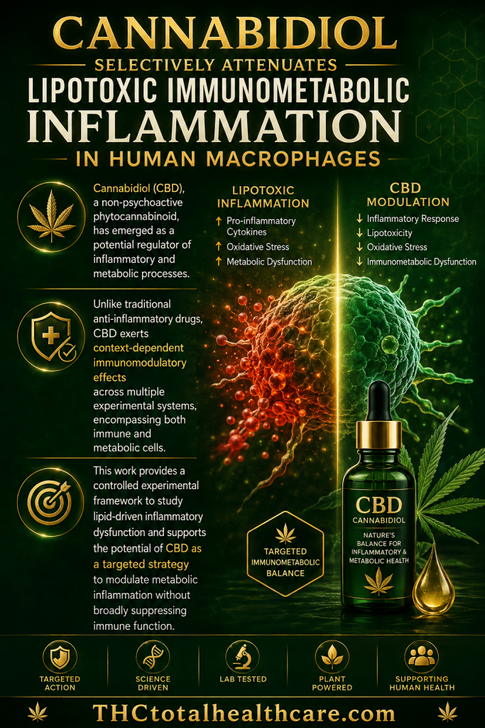
Cannabidiol selectively attenuates lipotoxic immunometabolic inflammation in human macrophages

Lipotoxic inflammation caused by excess fatty acids can drive metabolic disease by altering immune-cell function. In human macrophages, cannabidiol (CBD) selectively reduced inflammatory and immunometabolic responses triggered by lipotoxic stress without broadly suppressing normal immune activity. The researchers concluded that CBD may have therapeutic potential for metabolic disorders characterized by chronic lipid-driven inflammation.

“The role of saturated fatty acid-induced immunometabolic stress in macrophage dysfunction during metabolic disease remains incompletely understood, particularly the interplay between inflammatory signaling and intracellular lipid handling.

We employed a tightly controlled palmitic acid (PA)-based lipotoxicity model in PMA-differentiated U937-derived human macrophage-like cells to investigate how lipid excess reshapes inflammatory responses and to evaluate the modulatory effects of cannabidiol (CBD).

PA exposure induced a metabolically stressed yet viable macrophage phenotype, characterized by a broad cytokine remodeling profile. This included induction of classical proinflammatory cytokines such as interleukin (IL)-6, together with activation of inflammasome-associated cytokines IL-1β and IL-18 and additional immunoregulatory mediators, while tumor necrosis factor alpha (TNF-α) contributed to the overall inflammatory profile in a multivariate analysis. These changes were accompanied by a significant, time-dependent storage of intracellular triglycerides (TG) consistent with lipid overload and altered lipid handling.

CBD co-treatment did not compromise cell viability but selectively attenuated PA-induced inflammatory response in a cytokine-dependent manner, with the most significant reduction observed at higher concentrations. In parallel, CBD significantly reduced intracellular TG accumulation under lipotoxic conditions.

Collectively, these findings define a lipotoxicity-associated macrophage phenotype driven by saturated fatty acids and identify CBD as a context-dependent modulator of immunometabolic inflammation.

This work provides a controlled experimental framework to study lipid-driven inflammatory dysfunction and supports the potential of CBD as a targeted strategy to modulate metabolic inflammation without broadly suppressing immune function.”

https://pubmed.ncbi.nlm.nih.gov/42534637

“Cannabidiol (CBD), a non-psychoactive phytocannabinoid, has emerged as a potential regulator of inflammatory and metabolic processes. Unlike traditional anti-inflammatory drugs, CBD exerts context-dependent immunomodulatory effects across multiple experimental systems, encompassing both immune and metabolic cells.”

“Overall, this work supports CBD as a context-dependent modulator of immunometabolism response in PMA-differentiated U937-derived macrophages and provides a controlled experimental framework for studying lipid-driven inflammatory dysfunction in U937-derived macrophages.”

https://www.frontiersin.org/journals/immunology/articles/10.3389/fimmu.2026.1873494/full

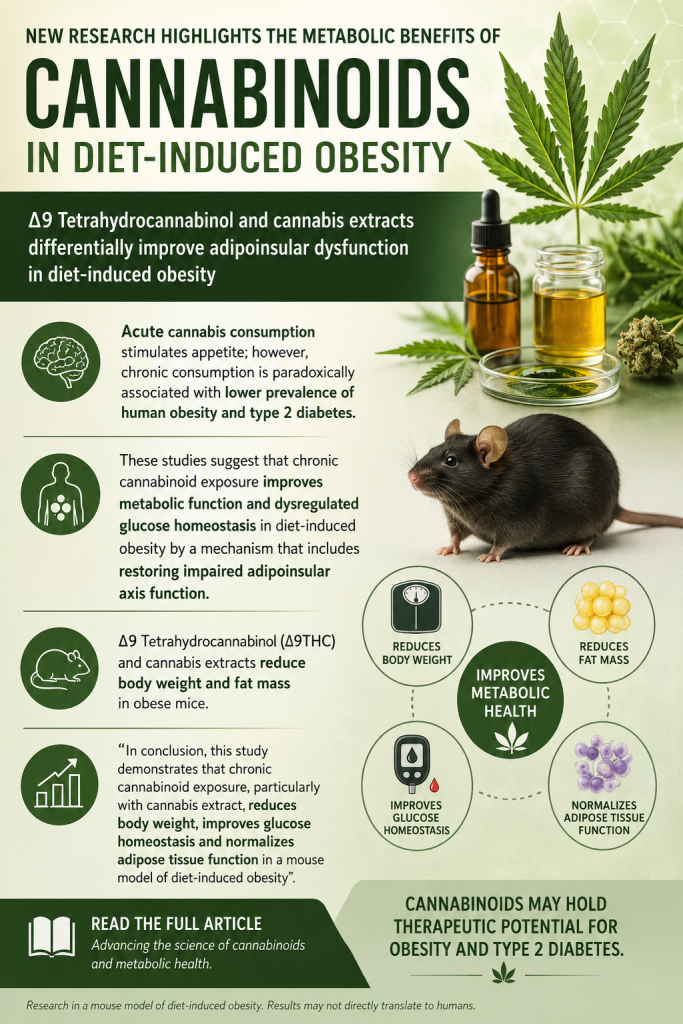
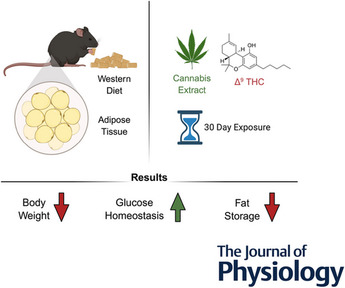
Δ9 Tetrahydrocannabinol and cannabis extracts differentially improve adipoinsular dysfunction in diet-induced obesity

THC and cannabis extracts may have important effects on obesity-related metabolic dysfunction. In this study, both THC and cannabis extracts reduced body weight and fat mass in diet-induced obese mice while improving abnormalities in adipose-tissue signaling. Cannabis extracts produced the strongest metabolic effects, more effectively normalizing adipokine expression and restoring glucose clearance to levels seen in lean mice, an effect not achieved by THC alone. The findings suggest that chronic cannabinoid exposure—particularly whole cannabis extracts—can improve glucose homeostasis, adipose function, and metabolic regulation in obesity.

“Diet-induced obesity (DIO) is associated with dysregulated adipoinsular axis and endocannabinoid system (eCBS) function. Acute cannabis consumption stimulates appetite; however, chronic consumption is paradoxically associated with lower prevalence of human obesity and type 2 diabetes.

We investigated the impact of chronic exposure to Δ9 tetrahydrocannabinol (THC) and cannabis extracts on DIO and glucose homeostasis in mice.

Male mice were fed a high-fat/sucrose diet or a low-fat/no-sucrose diet for 60 days. At day 30, mice were administered THC (5 mg/kg) or cannabis extracts matched for THC content daily for 30 days. We assessed adipocyte biology, glucose tolerance, insulin sensitivity, eCBS expression, body weight, food intake and motor activity. Roles for the eCBS in cannabis-induced changes in metabolic processes, including cellular bioenergetics, were analysed in 3T3-L1 adipocytes.

THC and extracts reduced body weight and fat mass in DIO mice, and reversed DIO-associated changes in expression of adipokines that regulate the adipoinsular axis. Extracts normalized expression of adipokines more effectively than THC. Notably, extracts – but not THC – normalized glucose clearance in DIO mice to levels found in lean mice. In addition, THC and extracts promoted anti-adipogenic effects and changes in energy metabolism in 3T3-L1 cells in a concentration-dependent manner.

These studies suggest that chronic cannabinoid exposure improves metabolic function and dysregulated glucose homeostasis in DIO by a mechanism that includes restoring impaired adipoinsular axis function.

KEY POINTS: Δ9 Tetrahydrocannabinol (Δ9THC) and cannabis extracts reduce body weight and fat mass in obese mice. Cannabis extracts, but not Δ9THC alone, improve glucose homeostasis in obese mice. Extracts more effectively normalize expression of components of the adipoinsular axis in obese mice. Δ9THC and extracts promote anti-adipogenic effects in 3T3-L1 cells. Δ9THC and extracts alter cellular bioenergetics in 3T3-L1 cells.”

https://pubmed.ncbi.nlm.nih.gov/42113966

“In conclusion, this study demonstrates that chronic cannabinoid exposure, particularly with cannabis extract, reduces body weight, improves glucose homeostasis and normalizes adipose tissue function in a mouse model of DIO. Our findings highlight the potential therapeutic value of cannabinoids in managing obesity and related metabolic disorders, though further research is needed to fully understand the underlying mechanisms and translate these findings into clinical applications.”

https://physoc.onlinelibrary.wiley.com/doi/10.1113/JP290431

Comparative Anti-Obesity Potential of Cannabigerol-Dominant Cannabis sativa L. Inflorescence Extracts via Differential Regulation of Lipid Metabolism in 3T3-L1 Cells

“Obesity is a chronic metabolic disorder characterized by excessive accumulation of body fat and is a major risk factor for various diseases, including type 2 diabetes, hypertension, and cardiovascular diseases.

This study investigated the anti-obesity effects of cannabigerol-dominant C. sativa inflorescence extracts (CEs) obtained using various ethanol concentrations.

The extracts were analyzed by UPLC to determine their major components. Additionally, anti-obesity mechanisms of the extracts were further determined through RT-qPCR and Western blot analysis to evaluate gene and protein expression levels. A total of seven cannabinoids, including cannabigerol as a major constituent, were identified within CE.

Differentiation of 3T3-L1 cells was dose-dependently inhibited by CE at all ethanol concentrations. Furthermore, the gene and protein expression levels of key adipogenic and lipogenic markers, such as PPARγ, C/EBPα, SREBP-1c, and FAS, were significantly downregulated by CE treatment. In contrast, the expression of factors involved in lipolysis and white adipose tissue browning, such as HSL, ATGL, UCP1, and PGC-1α, was markedly increased by CE treatment. These effects were enhanced in an ethanol concentration-dependent manner.

In conclusion, these results demonstrate that cannabigerol-dominant C. sativa effectively mitigates obesity by suppressing adipogenesis and lipogenesis while concurrently stimulating lipolysis and white adipose tissue browning.”

https://pubmed.ncbi.nlm.nih.gov/41751885

 “In conclusion, these results suggest that CE acts as a safe and effective therapeutic agent by simultaneously regulating adipogenesis, lipogenesis, lipolysis, and WAT browning.”

https://www.mdpi.com/1422-0067/27/4/1747


Aerobic training and cannabidiol activate the PI3K/AKT/PDX1 axis to ameliorate beta-cell dysfunction in a rat model of diet-induced obesity

Background: This study investigated the therapeutic potential of cannabidiol (CBD) and aerobic training (AT), both alone and in combination, to ameliorate beta-cell dysfunction in a rat model of diet-induced obesity, with a specific focus on the phosphatidylinositol 3-kinase (PI3K)/ protein Kinase B (AKT)/pancreatic and duodenal homeobox 1 (PDX1) pathway.

Methods: Thirty-two male Wistar rats were fed a high-fat diet (HFD) for 8 weeks to induce obesity. They were then randomly assigned to four groups (n = 8/group): HFD (sedentary), HFD + CBD (10 mg/kg, 5x/week), HFD + AT (30-minute treadmill running,50-80% maximal speed, 5x/week, 8 week), and HFD + CBD+AT (combined treatment) for a further 8 weeks. Following the intervention, beta-cell function was assessed via the HOMA-Beta index, and pancreatic gene expression of PI3K, AKT, and PDX1 was analyzed using RT-PCR.

Results: Both the HFD + CBD and HFD + CBD+AT groups showed a significant improvement in beta-cell function, as indicated by a higher HOMA-Beta index compared to the HFD group (p = 0.002 and p = 0.001, respectively). AT alone (HFD + AT) did not significantly alter HOMA-Beta. In contrast, all intervention groups (HFD + CBD, HFD + AT, and HFD + CBD+AT) demonstrated a significant upregulation in the gene expression of PI3K, AKT, and PDX1 compared to the HFD group (p < 0.001 for all). Notably, the combined treatment of CBD and AT (HFD + CBD+AT) produced a synergistic effect, resulting in a greater increase in the expression of all three genes compared to either intervention alone. No significant correlation was found between HOMA-Beta and the gene expression levels within any group (p > 0.05).

Conclusions: CBD and AT independently activate the pancreatic PI3K/AKT/PDX1 pathway, with their combination showing synergy. CBD, but not AT alone, improved functional beta-cell mass. This pathway activation represents a key mechanism for protecting beta-cells in obesity.”

https://pubmed.ncbi.nlm.nih.gov/41721911

https://link.springer.com/article/10.1007/s11033-026-11573-9

Cannabidiol mitigates high-fat-diet-induced early-stage inflammation in two adipose tissue fat depots of Wistar rats

“Cannabidiol (CBD) has potential for treating obesity-induced inflammation; thus, we studied the influence of CBD on the accumulation of lipid precursors of inflammation, the, enzymes, and cytokine levels in the subcutaneous (SAT) and visceral adipose tissue (VAT) of animals with obesity-induced early-stage inflammation.

Our experiment was performed on rats fed a high-fat (HFD) or control diet, which received CBD or its vehicle. The accumulation and composition of lipid fractions were assessed via gas‒liquid chromatography, whereas the expression of inflammatory pathway enzymes and the cytokine content were evaluated via Western blot or multiplexing, respectively.

In addition to selective changes in the content of cytokines, the administration of CBD to HFD-fed rats also decreased the deposition of all the lipid fractions in VAT, whereas in SAT, only the free fatty acid and diacylglycerol fractions were affected. Moreover, CBD reduced the deposition of arachidonic acid and the expression of enzymes associated with the synthesis of lipid precursors of inflammation in both the SAT and VAT of HFD-fed rats.

Although the data revealed the beneficial influence of CBD on lipid precursors of inflammation metabolism in both fat depots, more pronounced changes were observed in VAT, which is a tissue that is more predisposed to metabolic disease development.”

https://pubmed.ncbi.nlm.nih.gov/41571764

https://www.nature.com/articles/s41598-026-36666-0

Adipogenicity-induced human mesenchymal stem cells treated with hemp seed oil stimulate brown-like adipocytes and decrease adipokine levels through the activation of cannabinoid receptor 2 (CB2)

pubmed logo

“The endocannabinoid system (ECS) is essential for energy hemostasis, obesity, and other metabolic disorders.

Cannabidiol and polyunsaturated fatty acids (PUFAs), which are found in hemp seed oil (HSO), have been found to regulate adipose tissue through the ECS. Thus, human mesenchymal stem cells (hMSCs) were differentiated into pre-adipocytes and then treated with cannabidiol (CBD), tetrahydrocannabinol (THC), 0.05% HSO, or 0.1% HSO for 3 days (72 h).

The mixture was subsequently maintained in maintenance media for 14 days, after which the condition media (CM) was collected. In addition, THP-1 cells were used to assess the inflammatory response upon exposure to CM collected from different groups of experimental cells. Quantification for lipid accumulation (Oil red O), gene expression (RT‒qPCR), and protein levels (Western blot) were performed.

We found that HSO-treated cells matured toward brown-like adipose tissue with a spindle shape and decreased intracellular lipid accumulation. HSO treatment decreased the expression of genes associated with fat accumulation and browning (BAT), with the exception of UCP-1, which leans toward brown-like adipocytes. HSO treatment upregulated the cannabinoid receptors 2 (CB2), TRPV1, and GPCR55 mRNAs and leptin mRNA found with lower expression; no alterations were observed in cannabinoid receptors 1 (CB1), FAAH, and MGL mRNAs. In THP-1 macrophage, HSO treated CM decreased the expression of IL-6, IL-8, TNF-α, and leptin mRNAs significantly when compared to CBD and THC.

The potential of HSO in promoting brown fat characteristics through the CB2 and its effect on inflammation status offers an intriguing area for future research and therapeutic interventions.”

https://pubmed.ncbi.nlm.nih.gov/41258240

“Overall, the availability of balanced ratios of omega 3/omega 6 PUFAs and CBD in HSO favors in maintaining optimal ECS ligands in adipocytes. Our current study revealed that HSO treatment might promote the maturation of hMSC preadipocytes toward brown-like adipose tissue, which evident morphologically. ECS might mediate this effect, as HSO treatment downregulates the CB1 receptor and increases the CB2 receptor at the mRNA and protein levels. In addition, HSO treatment decreased inflammatory marker of IL-6, IL-8, TNF-α, and leptin compared to untreated cells; however, HSO treatment resulted in a minimalized the provoking of inflammatory cytokines compared with CBD and THC treatments in THP-1 cells. In conclusion, the potential of HSO in promoting the development of brown fat characteristics through the ECS and its effect on inflammation status offers an intriguing area for future research and therapeutic interventions.”

https://jcannabisresearch.biomedcentral.com/articles/10.1186/s42238-025-00343-2