Cannabinoids in Cancer: Molecular Mechanisms of Tumor Cell Death and Translational Opportunities

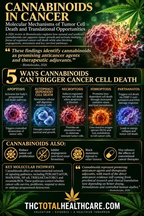
Cannabinoids can attack cancer cells through far more than a single mechanism. This 2026 review describes how natural and synthetic cannabinoids can suppress tumor-cell growth, angiogenesis and metastasis while activating multiple forms of regulated cancer-cell death, including apoptosis, autophagy-dependent cell death, necroptosis, ferroptosis and parthanatos.

The researchers detail an interconnected network of molecular pathways affected by cannabinoids, including PI3K/AKT/mTOR, MAPK/ERK, NF-κB, TRIB3/AKT/mTORC1 and ceramide/ROS signaling. These pathways control whether cancer cells survive, proliferate, respond to stress or undergo programmed destruction, giving cannabinoids multiple potential points of attack against malignant cells.

The review also finds that cannabinoids may enhance conventional anticancer treatments by engaging complementary cell-death pathways. The authors conclude that cannabinoids represent promising anticancer agents and therapeutic adjuvants, with much of the direct anticancer evidence still coming from preclinical research and clinical translation now depending on better dosing, formulations and controlled human studies.

“Cannabinoids are terpenophenolic compounds derived from Cannabis sativa L. that exert a broad range of biological and pharmacological activities. Increasing evidence highlights their potential as modulators of cancer progression specifically through the suppression of tumor cell growth, angiogenesis, and metastasis across multiple tumor models.

This review provides a comprehensive overview of the molecular mechanisms by which natural and synthetic cannabinoids induce regulated cancer cell death.

Current evidence demonstrates that cannabinoids regulate multiple forms of cancer cell death, including apoptosis, autophagy-dependent cell death, necroptosis, ferroptosis, and parthanatos. These effects are mediated through complex and interconnected signaling pathways such as TRIB3/AKT/mTORC1, PI3K/AKT/mTOR, MAPK/ERK, NF-κB, ERK/JNK/p38-MAPK, and ceramide/Raf1/ERK/ROS.

In addition to their direct antitumor effects, cannabinoids can enhance the efficacy of conventional anticancer therapies through the coordinated regulation of complementary cell death pathways. They also provide clinically relevant supportive benefits in palliative care, alleviating chemotherapy-induced nausea, cachexia, and mood or sleep disturbances.

Collectively, these findings identify cannabinoids as promising anticancer agents and therapeutic adjuvants, predominantly in the preclinical setting. However, significant challenges remain regarding their safety, optimal dosing, formulation, and clinical efficacy. Further mechanistic studies, rigorous preclinical research, and well-designed clinical trials are required to establish the translation of cannabinoid-based therapies into precision oncology.”

https://pubmed.ncbi.nlm.nih.gov/42793092

“The cannabis plant, a member of the Cannabaceae family, has been traditionally utilized for centuries for spiritual and remedial healing.”

“The anticancer activity of cannabinoids was discovered in 1975 by Munson et al.. Cannabinoids suppress cancer cell proliferation, angiogenesis, tumor invasion, metastasis, and stemness while inducing different modes of cell death pathways.”

“In summary, this review highlights the growing body of evidence supporting the ability of cannabinoids to induce multiple forms of cell death across a broad spectrum of cancer types. Through the modulation of diverse molecular and signaling pathways, cannabinoids have emerged as promising anticancer agents, mostly in preclinical models, with the potential to target tumor growth, survival, and progression, ultimately triggering diverse cell death mechanisms.”
https://www.mdpi.com/2218-273X/16/9/1260

Delta9-tetrahydrocannabinol induces apoptosis in C6 glioma cells

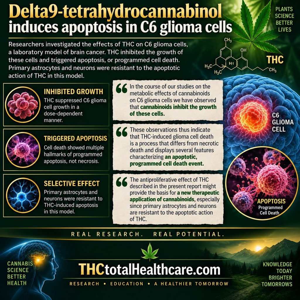
Researchers investigated whether Δ9-tetrahydrocannabinol (THC) could directly affect the survival of C6 glioma cells, a laboratory model of brain cancer. THC produced a dramatic, dose-dependent decline in cell metabolism and triggered apoptosis, or programmed cell death. The researchers confirmed this through several markers, including loss of membrane symmetry and the characteristic fragmentation of DNA seen during apoptosis.

The study also found that THC stimulated sphingomyelin breakdown, a process linked to ceramide signaling and cell death. Importantly, THC induced apoptosis in several transformed neural cell types but did not produce the same apoptotic effect in primary astrocytes or neurons. Even though the glioma cells expressed CB1 receptors, blocking CB1 did not prevent THC-induced apoptosis, suggesting that the effect occurred through a CB1 receptor-independent mechanism.

The authors concluded that THC’s antiproliferative effect could provide a basis for exploring new therapeutic applications of cannabinoids, particularly because normal primary astrocytes and neurons were resistant to THC-induced apoptosis in this model.

“delta9-Tetrahydrocannabinol (THC), the major active component of marijuana, induced apoptosis in C6.9 glioma cells, as determined by DNA fragmentation and loss of plasma membrane asymmetry. THC stimulated sphingomyelin hydrolysis in C6.9 glioma cells. THC and N-acetylsphingosine, a cell-permeable ceramide analog, induced apoptosis in several transformed neural cells but not in primary astrocytes or neurons. Although glioma C6.9 cells expressed the CBI cannabinoid receptor, neither THC-induced apoptosis nor THC-induced sphingomyelin breakdown were prevented by SR141716, a specific antagonist of that receptor. Results thus show that THC-induced apoptosis in glioma C6.9 cells may rely on a CBI receptor-independent stimulation of sphingomyelin breakdown.”

https://pubmed.ncbi.nlm.nih.gov/9771884

“In the course of our studies on the metabolic effects of cannabinoids on C6 glioma cells we have observed that cannabinoids inhibit the growth of these cells.”

“The antiproliferative effect of THC described in the present report might provide the basis for a new therapeutic application of cannabinoids, especially since primary astrocytes and neurons are resistant to the apoptotic action of THC.”

“These observations thus indicate that THC-induced glioma cell death is a process that differs from necrotic death and displays several features characterizing an apoptotic, programmed cell death event.”

https://febs.onlinelibrary.wiley.com/doi/10.1016/S0014-5793%2898%2901085-0

Need this THC study explained? thcXplained.com

Cannabidiol Suppresses Glioma Growth and Limits Invasion Partly Through an LOXL2-Associated EMT-Like Program

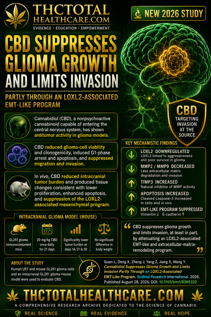
A new study found that cannabidiol (CBD) suppressed glioma growth and reduced tumor-cell migration and invasion in laboratory and animal models. The researchers linked part of this effect to reduced LOXL2 activity, suggesting CBD may interfere with molecular programs that help glioma cells spread into surrounding brain tissue.

The study adds new mechanistic evidence to the growing body of research examining CBD as a potential therapeutic compound in glioma and glioblastoma.

“Background: Gliomas, particularly glioblastoma, remain difficult to control because diffuse infiltration into surrounding brain tissue limits complete resection and contributes to recurrence. Cannabidiol (CBD), a nonpsychoactive cannabinoid capable of entering the central nervous system, has shown antitumor activity in glioma models, but the mechanisms underlying its anti-invasive effects remain unclear. Lysyl oxidase-like 2 (LOXL2) regulates extracellular-matrix remodeling and mesenchymal phenotypes in several cancers. We therefore tested the hypothesis that CBD limits glioma growth and invasion partly by suppressing an LOXL2-associated extracellular-matrix and EMT-like program.

Methods: Human U87 and murine GL261 glioma cells were used to examine CBD effects on tetrazolium-based cell viability, clonogenic growth, cell-cycle progression, apoptosis, migration, and invasion. The two cell lines provided complementary human and murine models, and the immunocompetent intracranial GL261 model enabled syngeneic in vivo validation. RNA sequencing and public glioma datasets were used to identify and contextualize CBD-responsive molecules. Mechanistic involvement was tested by determining whether LOXL2 knockdown phenocopied and LOXL2 overexpression attenuated the anti-invasive effects of CBD.

Results: CBD reduced glioma-cell viability and clonogenicity, induced G1-phase arrest and apoptosis, and suppressed migration and invasion. C CCK-8-derived IC50 values (mean ± SD, n = 3) at 24, 48, and 72 h were 36.5 ± 0.3, 26.7 ± 0.3, and 21.4 ± 0.3 μM in U87 cells and 33.3 ± 0.2, 29.3 ± 0.2, and 25.5 ± 0.4 μM in GL261 cells, respectively. CBD treatment was accompanied by reduced MMP2 and MMP9 expression and increased TIMP3 expression. Transcriptomic profiling identified LOXL2 as a prominent CBD-downregulated molecule, and public datasets associated higher LOXL2 expression with aggressive molecular features and shorter overall survival. LOXL2 silencing reproduced the antimigratory and anti-invasive phenotype, whereas LOXL2 overexpression enhanced cell motility and partially attenuated the effects of CBD. The partial rescue involved vimentin, MMP9/TIMP3, EMT-related transcription factors, and F-actin-rich protrusions. In vivo, CBD reduced intracranial tumor burden and produced tissue changes consistent with lower proliferation, enhanced apoptosis, and suppression of the LOXL2-associated mesenchymal program.

Conclusions: CBD suppresses glioma growth and limits invasion, at least in part, by attenuating an LOXL2-associated EMT-like and extracellular-matrix-remodeling program. Because LOXL2 overexpression produced only a partial rescue and direct target engagement was not tested, LOXL2 should be interpreted as a functional mediator rather than the sole or direct molecular target of CBD. These findings support further validation in patient-derived and pharmacokinetically informed glioma models.”

https://pubmed.ncbi.nlm.nih.gov/42661582

https://onlinelibrary.wiley.com/doi/10.1155/bmri/6385332

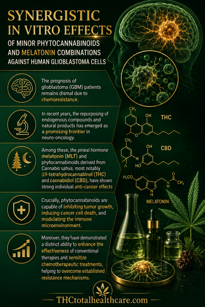
Synergistic In Vitro Effects of Minor Phytocannabinoids and Melatonin Combinations Against Human Glioblastoma Cells

Glioblastoma remains one of the most difficult brain cancers to treat, in part because tumor cells can develop resistance to chemotherapy. In laboratory testing, combinations of the minor cannabinoids CBN or CBG with melatonin produced synergistic anticancer effects against human glioblastoma cells while sparing healthy astrocytes, and significantly enhanced the effectiveness of temozolomide.

“The prognosis of glioblastoma (GBM) patients remains dismal due to chemoresistance.

Repurposing of natural and endogenous compounds, such as the pineal hormone melatonin (MLT) and minor phytocannabinoids like cannabinol (CBN) or cannabigerol (CBG), represents a promising strategy.

This study investigates the cytotoxic potential of combining these phytocannabinoids with MLT, evaluating their efficacy both alone and synergistically with temozolomide (TMZ) to overcome drug resistance.

To achieve this, cytotoxicity, synergy (Bliss model), and selectivity were evaluated in U87, T98, and U251 GBM lines and normal astrocytes. Mechanisms of damage were characterized via Western blot (γH2AX and PARP-1), flow cytometry using fluorescent dyes/probes (DCFDA, JC-1, MitoBright, BODIPY, PI, and Annexin-V), or the protein marker COX IV and confocal analysis.

The results demonstrated that CBN-MLT and CBG-MLT regimens exerted synergistic cytotoxicity while sparing healthy astrocytes. Notably, combining these regimens (U87: MLT 0.3 mg/mL + CBN 25 µM; MLT 0.2 mg/mL + CBG 15 µM. T98: MLT 0.7 mg/mL + CBN 25 µM; MLT 0.6 mg/mL + CBG 30 µM. U251: MLT 0.4 mg/mL + CBN 20 µM; MLT 0.5 mg/mL + CBG 35 µM) with TMZ significantly enhanced chemotherapeutic efficacy, overcoming baseline effects of TMZ in these cell lines.

The combinations induced necrotic cell death characterized by severe double-strand DNA damage. This was driven by an early accumulation of intracellular ROS, which triggered mitochondrial depolarization, loss of organelle mass, and lipid peroxidation. CBN combinations consistently triggered more robust biochemical alterations than CBG-based treatments.

Taken together, this study provides a strong preclinical basis for utilizing minor cannabinoids combined with MLT in GBM management.

Crucially, this co-treatment emerges as a promising approach to potentiate TMZ efficacy, offering a novel and potentially effective therapeutic strategy to counter GBM resilience.”

https://pubmed.ncbi.nlm.nih.gov/42589431

“In recent years, the repurposing of endogenous compounds and natural products has emerged as a promising frontier in neuro-oncology. Among these, the pineal hormone melatonin (MLT) and phytocannabinoids derived from Cannabis sativa, most notably Δ9-tetrahydrocannabinol (THC) and cannabidiol (CBD), have shown strong individual anti-cancer effects “

“Crucially, phytocannabinoids are capable of inhibiting tumor growth, inducing cancer cell death, and modulating the immune microenvironment. Moreover, they have demonstrated a distinct ability to enhance the effectiveness of conventional therapies and sensitize chemotherapeutic treatments, helping to overcome established resistance mechanisms.”

https://www.mdpi.com/1422-0067/27/15/6774


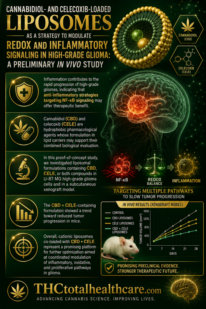
Cannabidiol- and Celecoxib-Loaded Liposomes as a Strategy to Modulate Redox and Inflammatory Signaling in High-Grade Glioma: A Preliminary In Vivo Study

High-grade glioma is marked by aggressive growth, inflammation, and oxidative stress, creating a need for therapies that can target several tumor-supporting pathways at once. In this preliminary in vivo study, liposomes carrying cannabidiol (CBD) and celecoxib modulated inflammatory and redox signaling in glioma tissue and showed antitumor effects. The researchers concluded that this combined liposomal approach may represent a promising strategy for further development against high-grade glioma.

“Inflammation contributes to the rapid progression of high-grade gliomas, indicating that anti-inflammatory strategies targeting NF-κB signaling may offer therapeutic benefit.

Cannabidiol (CBD) and celecoxib (CELE) are hydrophobic pharmacological agents whose formulation in lipid carriers may support their combined biological evaluation.

In this proof-of-concept study, we investigated liposomal formulations containing CBD, CELE, or both compounds in U-87 MG high-grade glioma cells and in a subcutaneous xenograft model.

We assessed cytotoxicity, apoptosis, oxidative stress, Nrf2-dependent responses, NF-κB-centered inflammatory networks, tumor cell invasive properties, and Wnt/β-catenin pathway activity. The nanoformulations induced reactive oxygen species generation by 1.8-fold, which was accompanied by Nrf2 activation. Cationic formulations loaded with the compounds produced more pronounced pro-apoptotic effects (up to 39%) than POPC liposomes, although both types reduced the nuclear translocation of the NF-κB p65 subunit.

The CBD + CELE-containing formulation showed a trend toward reduced tumor progression in mice. It is important to note that the in vitro and in vivo nanoformulations were physicochemically related, but not identical, and the in vivo experiment should be interpreted as a preliminary assessment after intratumoral administration.

Overall, cationic liposomes co-loaded with CBD + CELE represent a promising platform for further optimization aimed at coordinated modulation of inflammatory, oxidative, and proliferative pathways in glioma. However, additional studies, including tissue distribution, release kinetics, and efficacy in orthotopic glioma models, are needed to fully verify their translational potential.”

https://pubmed.ncbi.nlm.nih.gov/42511566

“In summary, this study shows that liposomal formulations containing CBD and CELE, particularly DOTAP:POPC formulations prepared with both compounds, modulate apoptosis, cell cycle distribution, oxidative stress, Nrf2/NF-κB/Wnt-related pathways, and glioma-associated inflammatory mediators in U-87 MG-based models. In the subcutaneous xenograft model, intratumoral administration of the CBD + CELE-containing formulation was associated with a trend toward reduced tumor progression and modulation of selected tumor-associated proteins.”

https://www.mdpi.com/1422-0067/27/14/6220

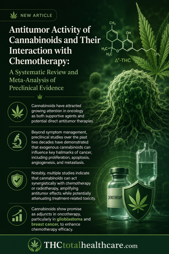
Antitumor Activity of Cannabinoids and Their Interaction with Chemotherapy: A Systematic Review and Meta-Analysis of Preclinical Evidence

Cannabinoids are increasingly being studied for direct antitumor effects and for their ability to influence the response to conventional chemotherapy. This systematic review and meta-analysis of preclinical research found that cannabinoids significantly reduced tumor growth and cancer-cell viability across multiple models. The analysis also identified evidence that cannabinoids can enhance the effectiveness of several chemotherapy agents, supporting the possibility of useful combination strategies. The findings strengthen the case for cannabinoids as biologically active anticancer compounds with potential both as direct therapies and as adjuncts to standard treatment.

“Background: Cannabinoids are studied as anticancer agents, but their effects vary across tumors, compounds, and experimental settings, underscoring the need to define consistent patterns. Our objective was to map cannabinoid efficacy across cancer preclinical models and identify tumor settings with the greatest translational promise. 

Methods: The protocol was registered on PROSPERO (CRD42025543744); PubMed, Embase, and CENTRAL were searched on 4 April 2024 for in vitro and in vivo studies assessing cannabinoid antitumor effects alone or with chemotherapy versus vehicle or chemotherapy only. Random-effects models yielded pooled mean differences (MD) with 95% confidence intervals (CI). MDs of viable cells were calculated for in vitro assays and tumor volume (mm3) for in vivo studies. Reports of various compounds, cannabidiol (CBD), tetrahydrocannabinol (THC) or synthetic cannabinoids, were pooled. 

Results: We included 189 studies in the final analysis. In vitro, cannabinoids reduced cell viability modestly overall, with significant effects in glioblastoma (MD -18.77 [CI: -27.15; -10.39]) and a nonsignificant trend in breast cancer (MD -6.75 [CI: -13.90; 0.40]). For in vivo, monotherapy showed the most consistent efficacy in glioblastoma, significantly reducing tumor volume by MD -980.58 mm3; [CI: -1270.2; -690.88]. Addition to temozolomide produced a favorable but nonsignificant decrease of MD -220.65 mm3; [CI: -579.34; 138.03, vs. temozolomide]. In breast cancer, cannabinoids achieved smaller yet significant tumor reductions (MD -402.64 mm3); [CI: -671.84; -133.45]. Synthetic agents had the largest effect (MD -1295.19 mm3); [CI: -1664.33; -928.05] -CBD plus doxorubicin vs. doxorubicin). Lung cancer (MD -562.17 mm3); [CI: -693.99; -430.35] and prostate cancer (MD -1136.59 mm3); [95% CI: -1320.97; -952.21] also had a significant response, whereas colon, pancreatic, and hepatocellular carcinoma models showed inconsistent or null responses. 

Conclusions: Cannabinoids show promise as adjuncts in oncotherapy, particularly in glioblastoma and breast cancer, to enhance chemotherapy efficacy. These findings should be interpreted with caution given the high inter-study heterogeneity typical of preclinical research and should be considered hypothesis-generating, warranting further validation in standardized and clinically relevant models.”

https://pubmed.ncbi.nlm.nih.gov/42198443

“Cannabinoids have attracted growing attention in oncology as both supportive agents and potential direct antitumor therapies.”

“Beyond symptom management, preclinical studies over the past two decades have demonstrated that exogenous cannabinoids can influence key hallmarks of cancer, including proliferation, apoptosis, angiogenesis, and metastasis.”

“Notably, multiple studies indicate that cannabinoids can act synergistically with chemotherapy or radiotherapy, amplifying antitumor effects while potentially attenuating treatment-related toxicity. These interactions are clinically appealing, as they suggest a capacity to sensitize tumor cells to conventional agents and possibly enable dose reductions that limit systemic adverse effects.”

“Cannabinoids show emerging potential as adjuncts in oncological treatment, with relatively consistent signals observed particularly in glioblastoma and breast cancer models.”

https://www.mdpi.com/1424-8247/19/5/768

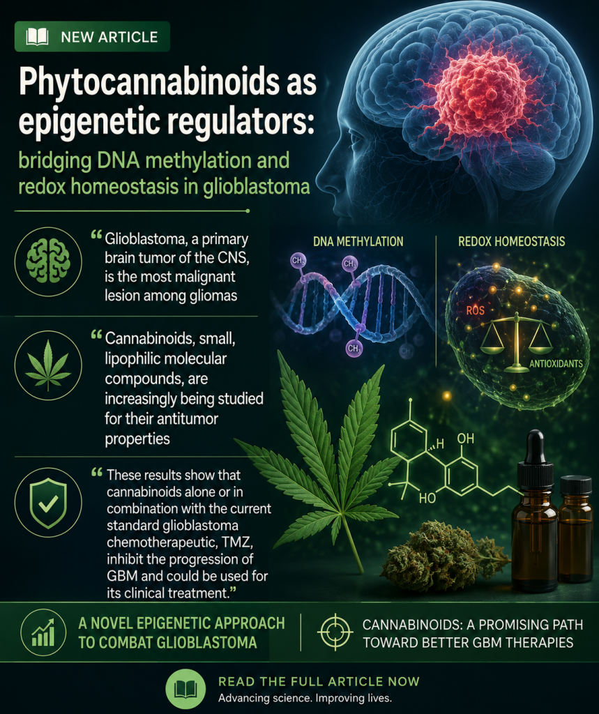
Phytocannabinoids as epigenetic regulators: bridging DNA methylation and redox homeostasis in glioblastoma

Glioblastoma is driven not only by genetic mutations, but also by epigenetic changes and oxidative stress that help tumor cells survive and resist treatment. This review examines how phytocannabinoids may influence both DNA methylation and redox balance in glioblastoma, potentially altering gene expression while disrupting cancer-cell defenses against oxidative damage. The evidence suggests that cannabinoids may act as epigenetic regulators as well as metabolic stressors, giving them multiple ways to interfere with glioblastoma progression and treatment resistance. The review highlights cannabinoid-based strategies as a promising direction for targeting the complex biology of this aggressive brain cancer.

“Glioblastoma, a primary brain tumor of the CNS, is the most malignant lesion among gliomas. It has a median survival time of about 12-15 months after diagnosis and limited treatment options.

That neoplastic processes result from changes in the cell’s redox potential and the overproduction of reactive oxygen species. As a consequence, the epigenetic marker, m5C of DNA, is oxidized with ROS to 5-hydroxymethylcytosine, but guanosine is damaged to 8-oxo-dG, a general probe of oxidative stress. If so, the m5C, as well as 8-oxo-dG content in DNA, are subject to dynamic changes induced by environmental and endogenous cellular factors. These markers can be used to evaluate new therapeutic agents, among others.

Currently, there are no effective drugs against human glioblastoma.

Cannabinoids, small, lipophilic molecular compounds, are increasingly being studied for their antitumor properties.

Using the precise nucleotide post-labelling method and thin-layer chromatographic analysis we monitored the effect of CBD, THC, and CFE, as well as their combination with temozolomide, on changes of global m5C and 8-oxo-dG contents.

These results show that cannabinoids alone or in combination with the current standard glioblastoma chemotherapeutic, TMZ, inhibit the progression of GBM and could be used for its clinical treatment. The mechanism of cannabinoids’ actions on glioblastoma cells is also proposed.”

https://pubmed.ncbi.nlm.nih.gov/42118523

“Recently, cannabinoids have gained attention for their anticancer properties. They are found, among others, in Cannabis sativa L.”

“This paper shows that CBD and THC induce hypermethylation and proposes a novel mechanism of action for cannabinoids in glioblastoma.”

https://link.springer.com/article/10.1007/s13353-026-01070-x

Acute anti-proliferative and anti-migratory effects of cannabidiol on C6 rat glioma, SH-SY5Y human neuroblastoma, and HT22 mouse hippocampal neuronal cell cultures

Cannabidiol continues to show direct effects on cancer-cell growth and movement across different tumor models. In this study, CBD reduced proliferation and migration in C6 glioma and SH-SY5Y neuroblastoma cells, demonstrating activity against two distinct types of neural cancer cells. The researchers also compared these effects with noncancerous HT22 hippocampal neuronal cells, helping clarify how CBD influences malignant versus normal neural tissue. The findings add to growing evidence that CBD may interfere with key processes involved in brain-tumor growth and spread.

“Background: The treatment of central nervous system tumors remains challenging owing to their highly proliferative nature, aggressiveness, and poor prognosis. Additionally, existing treatment methods have several problems, including high risk of complications, systemic side effects, and impact on patients’ quality of life. Recently, cannabidiol (CBD), a non-psychoactive cannabinoid found in Cannabis sativa, has emerged as an alternative therapeutic medication because of its potential antitumor activity with fewer side effects.

Methods: We evaluated the cell viability, clonogenicity, migration, apoptotic nuclear morphology, and cell cycle phases of C6 rat glioma, SH-SY5Y human neuroblastoma, and HT22 immortalized mouse hippocampus neuronal cultures treated with CBD ranged between 0 and 10 μg/mL.

Results: CBD concentrations exceeding 5 μg/mL induced significant reductions in cell viability in C6 glioma and SH-SY5Y neuroblastoma cultures, accompanied by decreased clonogenicity in both cultures at 10 μg/mL. A scratch assay for cell migration revealed that 5 μg/mL CBD suppressed C6 glioma cell migration. Additionally, late apoptotic nuclear morphology was observed in C6 glioma cultures treated with 10 μg/mL cannabidiol. Similarly, HT22 hippocampal neuronal cultures exhibited decreased cell viability and clonogenicity, with apparent nuclear signs of apoptosis at CBD concentrations over 5 μg/mL. Notably, CBD disrupted HT22 cell migration at concentrations of 2.5 and 5 μg/mL. Proteomic profiling of C6 glioma revealed upregulation of ribosomal proteins, molecular chaperones, and modulators of cytoskeletal dynamics upon treatment with 1 μg/mL CBD. In comparison, treatment with 2.5 μg/mL CBD led to marked downregulation of endoplasmic reticulum chaperones, mitochondrial ATP synthase, and cytoskeletal regulators.

Conclusion: Our findings confirm the sensitivity of glioma, neuroblastoma, and hippocampal neuronal cultures to CBD, providing valuable insights for further research into its therapeutic potential against glioma, neuroblastoma, and neuronal disorders.”

https://pubmed.ncbi.nlm.nih.gov/42088408

“Our findings demonstrate that CBD exerts dose-dependent anti-proliferative effects across all three cell lines, with tumor cells and neuronal cells exhibiting comparable sensitivity at higher concentrations.”

https://www.frontiersin.org/journals/toxicology/articles/10.3389/ftox.2026.1727831/full

The iron fist of nature: Cannabinoid derivatives alter iron homeostasis and activate ferroptotic pathways in glioblastoma cells

“Glioblastoma multiforme is the most commonly diagnosed type of brain tumor, with a poor prognosis and a high rate of recurrence. Because of its highly aggressive nature and the lack of efficient treatment options, novel therapeutic strategies are needed.

Ferroptosis is an iron-dependent, unique type of cell death, which provides an alternative way to eradicate cancer cells that are resistant to apoptosis and other cell death mechanisms.

CP55-940 (CP) and WIN 55212-2 (WIN) are synthetic cannabinoid receptor agonists with various biological activities, including neuroprotective and anticancer effects; however, their mechanism of action has not been fully uncovered.

In the present study, the potential of CP and WIN in glioblastoma cells was investigated.

Cell viability was determined with the MTT assay. Labile iron pool and reactive oxygen species generation were visualized with confocal microscopy. Malondialdehyde assay was performed to detect lipid peroxidation. Gene expressions of ferroptotic hallmarks, glutathione peroxidase-4, and transferrin receptor 1 were determined by RT-qPCR. Protein expression levels of iron-responsive element-binding protein 2, solute carrier family 7 member 11, and glutathione peroxidase-4 were analyzed by western blotting.

Results demonstrated that CP and WIN significantly induce ferroptotic pathways in glioblastoma cells via increased oxidative stress, labile iron pool, and lipid peroxidation. Furthermore, it was determined for the first time that both compounds significantly upregulate the transferrin receptor 1 gene expression.

In conclusion, the present study demonstrated for the first time that cannabinoid derivatives CP and WIN alter iron regulation and initiate ferroptosis in glioblastoma cells, rendering them potential candidates in therapy.

SIGNIFICANCE STATEMENT: We explored the ferroptotic activity of cannabinoid derivatives (CP and WIN) in glioblastoma cells for the first time. Additionally, we report for the first time that cannabinoid derivatives alter cellular iron levels, causing increased labile iron pool via upregulating the transferrin gene significantly.”

https://pubmed.ncbi.nlm.nih.gov/41962357

https://jpet.aspetjournals.org/article/S0022-3565(26)00518-5/abstract

“CP 55,940 is a synthetic cannabinoid which mimics the effects of naturally occurring THC (one of the psychoactive compounds found in cannabis). CP 55,940 was created by Pfizer in 1974 but was never marketed. It is currently used as a research tool to study the endocannabinoid system.”

“WIN 55,212-2 is a chemical described as an aminoalkylindole derivative, which produces effects similar to those of cannabinoids such as tetrahydrocannabinol (THC) but has an entirely different chemical structure.”

Hemp-Derived Extracellular Vesicles: A Novel Frontier in Nanomedicine and Therapeutics

“While mammalian-derived extracellular vesicles (EVs) face significant challenges in clinical translation due to scalability, cost, and safety, plant-derived EVs (PDEVs) have emerged as a promising alternative.

This review focuses on EVs derived from hemp (Cannabis sativa L.), or HEVs, a particularly compelling source that combines the general benefits of PDEVs, such as improved safety and scalability, with a unique, inherent therapeutic cargo.

HEVs are naturally enriched with a potent mix of cannabinoids, terpenes, and flavonoids, which may enhance therapeutic outcomes through synergistic interactions-a phenomenon known as the ‘entourage effect.’

Preclinical studies already demonstrate their potential, showing significant anti-cancer effects against aggressive tumors like glioblastoma, along with neuroprotective and anti-inflammatory properties.

However, the critical challenge hindering their clinical application is the lack of standardized, GMP (Good Manufacturing Practice)-compliant manufacturing protocols to address the inherent biochemical variability of the source material.

Overcoming these obstacles will be vital to unlocking the potential of HEVs as a novel, scalable frontier in nanomedicine.”

https://pubmed.ncbi.nlm.nih.gov/41787227

https://link.springer.com/article/10.1007/s40259-026-00766-0