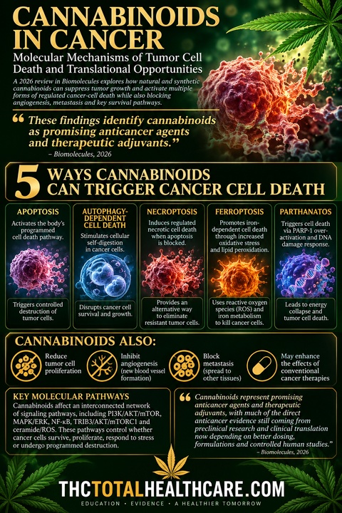
Cannabinoids can attack cancer cells through far more than a single mechanism. This 2026 review describes how natural and synthetic cannabinoids can suppress tumor-cell growth, angiogenesis and metastasis while activating multiple forms of regulated cancer-cell death, including apoptosis, autophagy-dependent cell death, necroptosis, ferroptosis and parthanatos.
The researchers detail an interconnected network of molecular pathways affected by cannabinoids, including PI3K/AKT/mTOR, MAPK/ERK, NF-κB, TRIB3/AKT/mTORC1 and ceramide/ROS signaling. These pathways control whether cancer cells survive, proliferate, respond to stress or undergo programmed destruction, giving cannabinoids multiple potential points of attack against malignant cells.
The review also finds that cannabinoids may enhance conventional anticancer treatments by engaging complementary cell-death pathways. The authors conclude that cannabinoids represent promising anticancer agents and therapeutic adjuvants, with much of the direct anticancer evidence still coming from preclinical research and clinical translation now depending on better dosing, formulations and controlled human studies.
“Cannabinoids are terpenophenolic compounds derived from Cannabis sativa L. that exert a broad range of biological and pharmacological activities. Increasing evidence highlights their potential as modulators of cancer progression specifically through the suppression of tumor cell growth, angiogenesis, and metastasis across multiple tumor models.
This review provides a comprehensive overview of the molecular mechanisms by which natural and synthetic cannabinoids induce regulated cancer cell death.
Current evidence demonstrates that cannabinoids regulate multiple forms of cancer cell death, including apoptosis, autophagy-dependent cell death, necroptosis, ferroptosis, and parthanatos. These effects are mediated through complex and interconnected signaling pathways such as TRIB3/AKT/mTORC1, PI3K/AKT/mTOR, MAPK/ERK, NF-κB, ERK/JNK/p38-MAPK, and ceramide/Raf1/ERK/ROS.
In addition to their direct antitumor effects, cannabinoids can enhance the efficacy of conventional anticancer therapies through the coordinated regulation of complementary cell death pathways. They also provide clinically relevant supportive benefits in palliative care, alleviating chemotherapy-induced nausea, cachexia, and mood or sleep disturbances.
Collectively, these findings identify cannabinoids as promising anticancer agents and therapeutic adjuvants, predominantly in the preclinical setting. However, significant challenges remain regarding their safety, optimal dosing, formulation, and clinical efficacy. Further mechanistic studies, rigorous preclinical research, and well-designed clinical trials are required to establish the translation of cannabinoid-based therapies into precision oncology.”
“The cannabis plant, a member of the Cannabaceae family, has been traditionally utilized for centuries for spiritual and remedial healing.”
“The anticancer activity of cannabinoids was discovered in 1975 by Munson et al.. Cannabinoids suppress cancer cell proliferation, angiogenesis, tumor invasion, metastasis, and stemness while inducing different modes of cell death pathways.”
“In summary, this review highlights the growing body of evidence supporting the ability of cannabinoids to induce multiple forms of cell death across a broad spectrum of cancer types. Through the modulation of diverse molecular and signaling pathways, cannabinoids have emerged as promising anticancer agents, mostly in preclinical models, with the potential to target tumor growth, survival, and progression, ultimately triggering diverse cell death mechanisms.”
The first time I heard Olivia Newton-John’s voice on the radio, I thought she might be an angel.
Decades later, when I heard that same voice on the news championing a controversial cause close to my heart, I felt I had been right.
In May 2017, before I knew her breast cancer had returned, I received an instant message from someone claiming to be Olivia Newton-John. The message encouraged me to keep doing what I was doing — sharing science — and echoed a phrase I had been posting for years:
“Thank you for sharing.”
I remember what happened next because I watched it unfold in real time.
The world learned that Olivia Newton-John’s breast cancer had returned and metastasized to her sacrum. By June, I was reading reports that she was using cannabis oil. By September, she was talking about cannabis openly.
And not timidly.
In an Australian 60 Minutes interview, Olivia called cannabis a “magical, miracle plant.” Days later, on American television, she called it “a healing plant.”
She said it helped her pain. She discussed inflammation. Over the following years, she would talk about cannabis helping her sleep and anxiety. She credited her husband, John Easterling, with growing cannabis and preparing tinctures specifically for her. She later said cannabis helped her wean herself off morphine.
“I couldn’t have got through this time without it,” she said in 2019.
Yet I remember watching another story develop alongside hers.
Olivia was telling the public how well she was doing, how important cannabis had become to her and how determined she was to live.
Much of the news coverage, however, increasingly felt like a countdown toward her death.
That became especially frustrating in 2018.
Olivia revealed that the cancer she had publicly disclosed in 2017 was actually her third experience with breast cancer. Her first diagnosis had come in 1992. She had experienced another recurrence in 2013 but chose to keep it private.
That was significant new information.
But it was not a new recurrence in 2018.
Nevertheless, headlines appeared announcing that Olivia Newton-John’s cancer had “returned for a third time.” To someone encountering the story casually, it could sound as though she had suffered another fresh setback.
The individual facts were technically true but the overall impression was misleading.
At the same time some headlines were emphasizing another “cancer battle,” Olivia was appearing publicly, participating in her Wellness Walk, talking about “winning over” cancer and telling interviewers she was doing well.
One source close to her pushed back against reports suggesting her condition had suddenly worsened, saying the information being reported was the same diagnosis she had disclosed the previous year.
I wanted to call attention to that disconnect then.
I did not have the ability to assemble years of interviews, headlines and contemporaneous reporting the way I can now.
But I remembered it.
And when the chronology is reconstructed, Olivia Newton-John’s story looks different.
She was never simply a celebrity who used marijuana.
Her relationship with cannabis became part of a much larger philosophy of cancer care.
Olivia had embraced complementary approaches since her first cancer diagnosis. She used conventional medicine, too. That point matters because her story is sometimes reduced to a false choice between cannabis and oncology.
Her approach was integrative.
Over the years she underwent surgery, chemotherapy, radiation, hormone therapy and other medical treatments while also using meditation, herbs, supplements, homeopathy, acupuncture, cannabis and other wellness practices.
She described treatment decisions as deeply personal.
Her husband, John Easterling, added another dimension.
Long before cannabis became central to their public story, Easterling had spent years studying plants and working with botanical medicine. Newton-John called him her “plant medicine man.”
The two seemed almost destined to find each other: a woman already drawn to holistic healing and a man who had devoted much of his life to medicinal plants.
By the time Newton-John was living with metastatic disease, Easterling was cultivating different cannabis strains at their California home and making tinctures for her.
Olivia said she did not like feeling intoxicated.
What she repeatedly emphasized was relief.
“If I don’t take the cannabis, I can feel the pain so I know it’s working,” she told 60 Minutes reporter Liz Hayes.
She called herself “incredibly pro cannabis.”
Her advocacy eventually moved beyond her own use.
“My dream is that it will be available to all the cancer patients or people going through cancer or any kind of disease that causes pain,” she said.
She talked about parents facing prosecution while seeking cannabis for children with epilepsy. She challenged the stigma surrounding the plant. She argued that patients deserved the right to try it.
And sometimes she went further than cautious symptom language.
She talked about cannabis and healing. She said research was examining the plant “not just for pain but to cure things.” She said she believed medicinal cannabis could play a major role in defeating cancer.
Those statements should be understood for what they were: the beliefs and hopes of a patient and advocate, not the results of a randomized clinical trial.
But that is precisely where her story becomes particularly important.
She did not stop at belief.
She wanted research.
That may be the part of her cannabis legacy most worth remembering.
Olivia spent years raising money and lending her name to what became the Olivia Newton-John Cancer Wellness & Research Centre in Melbourne, Australia. She insisted that “wellness” be part of the institution because she believed cancer care should treat the whole person, not merely the tumor.
Her later foundation explicitly sought to fund research into plant medicine.
And today, that idea is becoming reality.
The RESONANCE trial is studying medicinal cannabis in people with advanced cancer, examining symptoms, quality of life, tolerability and adverse effects.
The research represents something Olivia repeatedly called for: move beyond stigma, beyond anecdotes and beyond political arguments, and actually study what cannabis can and cannot do for patients.
That is why the timing of this story matters.
Olivia Newton-John would have turned 78 on September 26.
The medicinal-cannabis trial her advocacy helped inspire is actively recruiting.
October is Breast Cancer Awareness Month.
And on October 4, supporters will gather for the final Olivia’s Walk for Wellness.
The walk is ending.
The research is not.
Neither should our examination of the story Olivia Newton-John was trying to tell us.
For years, Olivia told interviewers that cannabis helped her through severe pain, improved her sleep, aided her quality of life and changed the way she thought about a plant she once approached nervously.
Then she used her celebrity to ask why other patients could not have the same opportunity.
Finally, she tried to move the conversation from testimony to action.
I watched her journey unfold online.
What frustrated me then was seeing a woman talking about healing, hope and the wonderful things she believed this plant was doing for her, while the public narrative so often seemed fixated on how close she was to dying.
What strikes me now that she is gone is what she left behind.
The headlines eventually stopped.
The interviews remain.
The cancer center remains.
The science continues.
Can cannabis fight cancer?
She believed it could.
Decades of laboratory and preclinical research have given scientists reason to keep asking the question Olivia Newton-John wanted answered.
The research she called for is being done.
Perhaps that is the most fitting way to remember her as her birthday approaches, as supporters prepare for the final Walk for Wellness.
Not merely as a celebrity who had cancer and used cannabis.
But as someone who turned what she learned from living with it for 30 years into a legacy that will walk on.
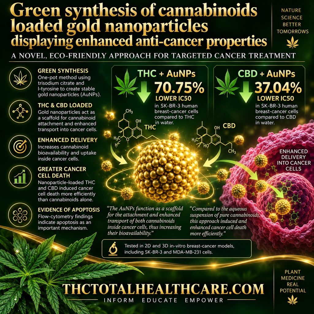
Researchers developed gold nanoparticles capable of carrying the highly hydrophobic cannabinoids THC and CBD into cancer cells, increasing their bioavailability and anti-cancer activity. The nanoparticles acted as a scaffold for cannabinoid attachment and enhanced transport inside cancer cells. Compared with aqueous suspensions of the pure cannabinoids, the nanoparticle-loaded THC and CBD produced cancer cell death more efficiently, supporting gold nanoparticles as a potential delivery system for cannabinoid-based cancer treatment.
“This study investigates the anti-cancer potential of cannabinoids loaded onto gold nanoparticles (AuNPs) for targeted cancer treatment. We adopted a multifaceted approach to explore and design this nano system, encompassing its synthesis, physicochemical characterization, stability assessment, and evaluation of anticancer efficacy in 2D and 3D in vitro models.
In this research, we have demonstrated that two highly hydrophobic phytocannabinoids like delta-9-tetrahydrocannabinol (THC) and cannabidiol (CBD), can be loaded on the surface of AuNPs with a one-pot synthesis protocol using trisodium citrate and l-tyrosine as a reducing and stabilizing agents. l-tyrosine plays a crucial role in cannabinoid loading, stability, and shelf life of the AuNPs.
After synthesis, the cannabinoid-loaded nanoparticles were characterized with UV–vis spectroscopy, dynamic light scattering (DLS), dark field hyperspectral microscopy, and electron microscopy. The AuNPs function as a scaffold for the attachment and enhanced transport of both cannabinoids inside cancer cells, thus increasing their bioavailability. Hyperspectral microscopy was used to confirm AuNPs uptake. IC50 values in SK-BR-3 human breast cancer cell line for both THC and CBD loaded onto AuNPs were lower by 70.75 % and 37.04 % than those of the aqueous suspension of pure molecules.
Compared to the aqueous suspension of pure cannabinoids, this approach induced and enhanced cancer cell death more efficiently. This enhanced efficacy was associated with a decline in cell viability, which is attributed to apoptosis, as indicated by flow cytometry results.
Our findings offer a significant step towards the green design and utilization of AuNPs to deliver cannabinoids into cells efficiently.”
“Cannabinoids loaded on AuNPs were synthesized as a nano-therapeutic drug agent for cancer. This study indicates that AuNPs can be used as efficient carriers for highly hydrophobic molecules with an anti-cancer effect. This protocol was straightforward and effective in entrapment of CBD or THC on the surface of AuNPs. The results revealed reasonable anti-cancer efficacy of this nanosystem in 2D and 3D in vitro cytotoxicity.”
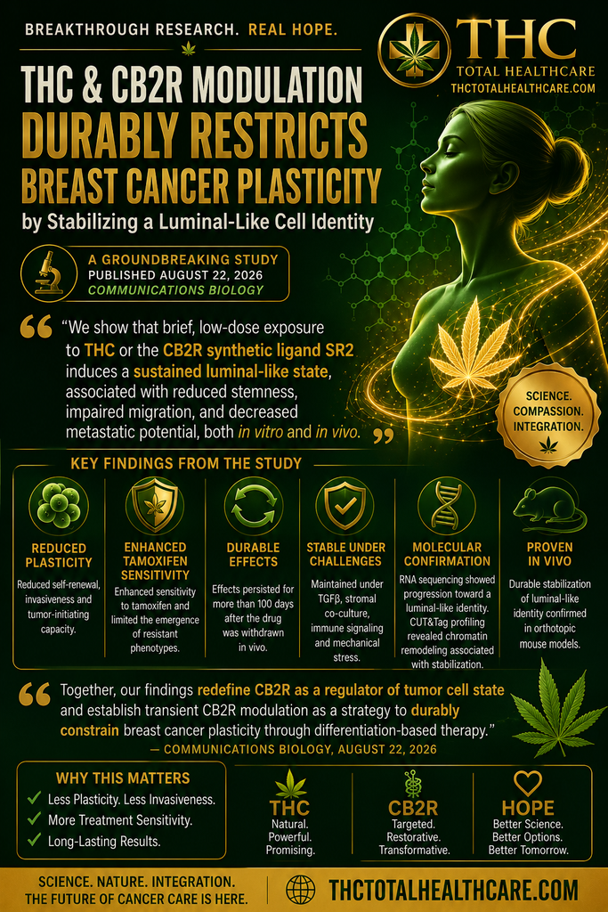
New breast cancer research suggests that brief modulation of cannabinoid receptor 2 (CB2R), including with THC, may have lasting effects on tumor cell behavior. Researchers found reduced self-renewal, invasiveness, and tumor-initiating capacity, along with greater tamoxifen sensitivity and a durable shift toward a more stable luminal-like cell identity.
“Cancer cell plasticity enables therapy resistance and metastasis by allowing transitions between stem-like and differentiated states. Differentiation-based strategies aim to stabilize tumor cell identity, yet pharmacological approaches that induce durable lineage restriction remain limited.
Here, we show that transient modulation of cannabinoid receptor 2 (CB2R) durably restricts breast cancer cell plasticity by stabilizing a luminal-like cell state. Using patient-derived and murine tumor organoids, we demonstrate that brief, low-dose CB2R modulation reduces self-renewal, invasiveness and tumor-initiating capacity, while enhancing tamoxifen sensitivity and limiting the emergence of resistant phenotypes.
These effects persist under pro-dedifferentiation conditions, including TGFβ exposure, stromal co-culture, immune signaling and mechanical stress, and are maintained in vivo following orthotopic transplantation. RNA sequencing reveals a progressive transition from an early plastic state toward a stabilized luminal-like identity, supported by CUT&Tag profiling that uncovers chromatin remodeling associated with this stabilization.
Together, our findings redefine CB2R as a regulator of tumor cell state and establish transient CB2R modulation as a strategy to durably constrain breast cancer plasticity through differentiation-based therapy.”
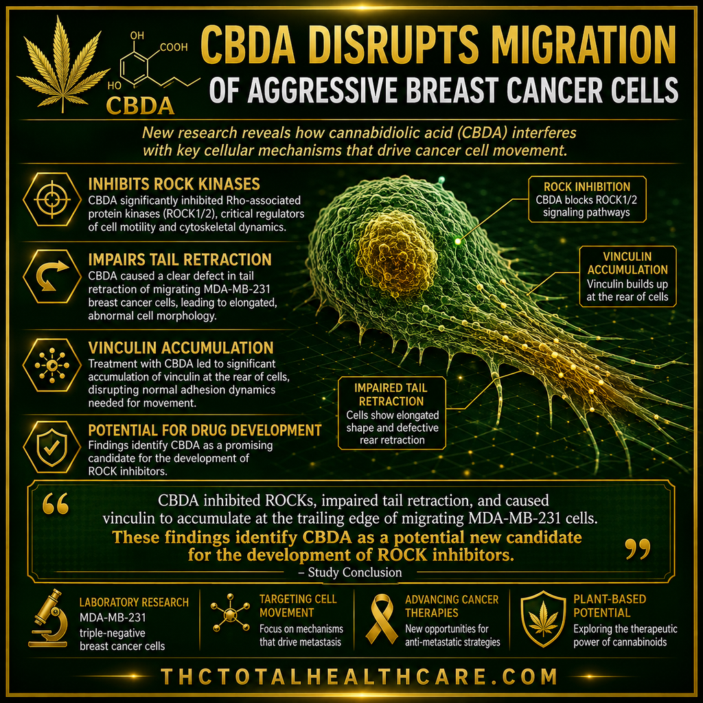
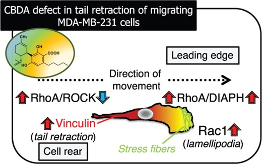
A new study adds mechanistic detail to earlier evidence that cannabidiolic acid (CBDA) can interfere with the migration of highly aggressive MDA-MB-231 breast cancer cells. Researchers found that CBDA inhibited Rho-associated kinases (ROCKs), impaired tail retraction, altered cell shape and caused vinculin to accumulate at the trailing edge—revealing another way this cannabis-derived compound may disrupt cellular behavior involved in cancer spread.
“We previously reported that cannabidiolic acid (CBDA), a major cannabinoid constituent of the fiber-type cannabis plant, abrogates the migration of highly aggressive human breast cancer MDA-MB-231 cells and activates the small GTPase RhoA by inhibiting protein kinase A. However, the mechanism(s) mediating RhoA signaling, which decreases cell migration, have not yet been comprehensively elucidated.
RhoA is an upstream mediator of Rho-associated kinases (ROCKs), diaphanous-related formins (DIAPHs), the RhoA-ROCK pathway (tail retraction) and the RhoA-DIAPH pathway (lamellipodia formation).
Herein, we identified CBDA as an inhibitor of ROCKs (at approximately 25 μM), which markedly elongated the cell body of MDA-MB-231 cells, similar to Y-27632, an established ROCK inhibitor.
CBDA stimulated lamellipodia formation at the leading edge, whereas NSC23766 (an established Rac1 inhibitor) completely blocked this elongated morphology. Biochemical analyses, including time-lapse imaging and confocal laser scanning microscopy, revealed that, compared to Y-27632, CBDA can induce impaired tail retraction coupled with unidirectional elongation of the cell body, upregulate the mRNA expression of DIAPHs and accumulate vinculin, an adhesion protein, at the trailing edge without affecting its expression.
These results indicate the potential of CBDA as a new candidate for the synthesis of ROCK inhibitors, which can evoke the directed elongation of MDA-MB-231 cells.”
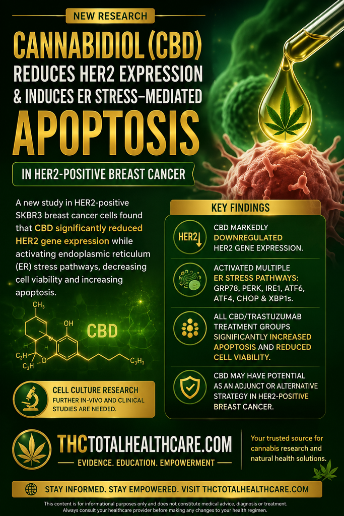
Researchers examining cannabidiol (CBD) in HER2-positive breast cancer cells found that CBD significantly reduced HER2 gene expression while activating endoplasmic reticulum (ER) stress pathways, decreasing cell viability, and increasing apoptosis. The findings identify another potential molecular mechanism behind CBD’s previously observed anticancer activity in breast cancer and suggest that CBD warrants further investigation both alone and in combination with HER2-targeted therapy.
“Cannabidiol (CBD) has attracted attention as a potential anticancer agent due to its observed efficacy, particularly in breast cancer. However, the exact molecular pathway through which it exerts its anticancer effects remains unknown. The present study aimed to investigate the effects of CBD on the HER2 receptor, endoplasmic reticulum (ER) stress signaling pathway, and apoptosis in HER2-positive breast cancer cells, as well as to examine the effect of trastuzumab (TZB), used in treatment, on the ER stress signaling pathway.
Methods
For this purpose, SKBR3 cells were treated with CBD at three different concentrations (9.38, 18.75, and 37.5 µM), TZB at 62.5 µg/mL, and two combinations of TZB and CBD (18.75 µM CBD + 62.5 µg/mL TZB and 37.5 µM CBD + 62.5 µg/mL TZB). Cell proliferation was assessed using real-time cell analysis, while expression levels of ER stress-associated genes were measured by qPCR. Apoptosis was analyzed by Annexin V/PI flow cytometry.
Results
Findings revealed that CBD, TZB, or their combination were associated with reduced cell proliferation, significant activation of ER stress pathways, and increased apoptosis, while CBD was associated with marked downregulation of HER2 gene expression. Treatment with 9.38 µM CBD, 18.75 µM CBD, 37.5 µM CBD, 62.5 µg/mL TZB, 18.75 µM CBD + 62.5 µg/mL TZB, and 37.5 µM CBD + 62.5 µg/mL TZB significantly increased GRP78, PERK, IRE1, ATF6, eIF2A, ATF4, CHOP, and XBP1s gene expression levels, and significantly decreased HER2 gene expression at all doses except 62.5 µg/mL TZB. All doses significantly increased the total number of apoptotic cells and were associated with significantly reduced cell viability.
Conclusions
The present findings demonstrated that both TZB and CBD are associated with activation of the ER stress response and induction of apoptosis in HER2-positive breast cancer cells, and that CBD is associated with downregulation of HER2 receptor gene expression. Collectively, our results indicate that CBD may have potential as an adjunct or alternative strategy in HER2-positive breast cancer, although further in vivo studies are required to evaluate the efficacy and safety of its combination with low-dose TZB.”
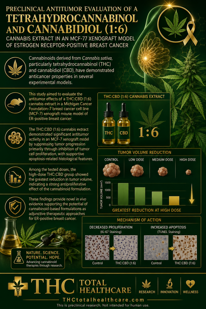
Estrogen receptor-positive breast cancer remains one of the most common forms of breast cancer, and new adjunctive treatment strategies are still needed. In this mouse xenograft study, a whole-cannabis extract containing THC and CBD in a 1:6 ratio significantly suppressed tumor growth, reduced tumor-cell proliferation, and increased apoptosis-related changes, with the highest dose producing the greatest reduction in tumor volume. The researchers concluded that the extract showed promising antiproliferative and antitumor activity and may warrant further investigation as an adjunctive approach for ER-positive breast cancer.
“Background and aim: Breast cancer remains one of the leading causes of cancer-related mortality worldwide, despite advances in surgery, chemotherapy, endocrine therapy, and targeted treatments. Cannabinoids derived from Cannabis sativa, particularly tetrahydrocannabinol (THC) and cannabidiol (CBD), have demonstrated anticancer properties in several experimental models; however, in vivo evidence in estrogen receptor (ER)-positive breast cancer remains limited. This study aimed to evaluate the antitumor effects of a THC:CBD (1:6) cannabis extract in a Michigan Cancer Foundation-7 breast cancer cell line (MCF-7) xenograft mouse model of ER-positive breast cancer.
Materials and methods: Female BALB/c nude mice bearing MCF-7 xenograft tumors were randomly assigned into five groups (n = 5/group): negative control (sesame oil), positive control treated with 5-fluorouracil (5-FU; 20 mg/kg), and three treatment groups receiving oral THC:CBD (1:6) extract at doses of 2, 10, or 20 mg/kg body weight for 30 consecutive days. Tumor growth was monitored throughout the experiment. Histopathological examination and immunohistochemical analysis of proliferating cell nuclear antigen (PCNA) expression were performed to evaluate apoptosis-related morphology and tumor cell proliferation. Hematological and biochemical parameters were assessed to determine systemic safety.
Results: Cannabinoid-treated groups exhibited significant suppression of tumor growth compared with the negative control group. Tumor volume reduction was observed in all treatment groups, with the greatest reduction detected in the high-dose THC:CBD group. Histopathological evaluation revealed increased numbers of tumor cells exhibiting morphological features consistent with apoptosis in cannabinoid-treated mice. Immunohistochemical analysis demonstrated significantly lower PCNA expression scores in all THC:CBD-treated groups compared with both negative and positive controls, indicating reduced tumor cell proliferation. Hematological parameters remained within normal physiological ranges in cannabinoid-treated animals. However, elevated alanine aminotransferase and aspartate aminotransferase levels were observed in the high-dose group, suggesting potential dose-related hepatic stress.
Conclusion: The THC:CBD (1:6) cannabis extract demonstrated significant antitumor activity in an MCF-7 xenograft model by suppressing tumor progression primarily through inhibition of tumor cell proliferation, with supportive apoptosis-related histological features. These findings provide novel in vivo evidence supporting the potential of cannabinoid-based formulations as adjunctive therapeutic approaches for ER-positive breast cancer.”
“In conclusion, the present study demonstrated that the THC:CBD (1:6) whole-cannabis extract exerted significant antitumor activity in an MCF-7 xenograft model of ER-positive breast cancer. Treatment with the cannabinoid extract resulted in marked suppression of tumor growth, significant reduction in tumor volume, decreased PCNA expression, and increased numbers of tumor cells exhibiting apoptosis-related morphological features.
Among the tested doses, the high-dose THC:CBD group showed the greatest reduction in tumor volume, indicating a strong antiproliferative effect of the cannabinoid formulation.”
“Overall, despite the inherent limitations of xenograft models, the present findings indicate that the THC:CBD (1:6) cannabis extract possesses promising antiproliferative and antitumor properties in ER-positive breast cancer. These results provide a foundation for future mechanistic and translational studies exploring cannabinoids as potential adjunctive agents in breast cancer therapy.”
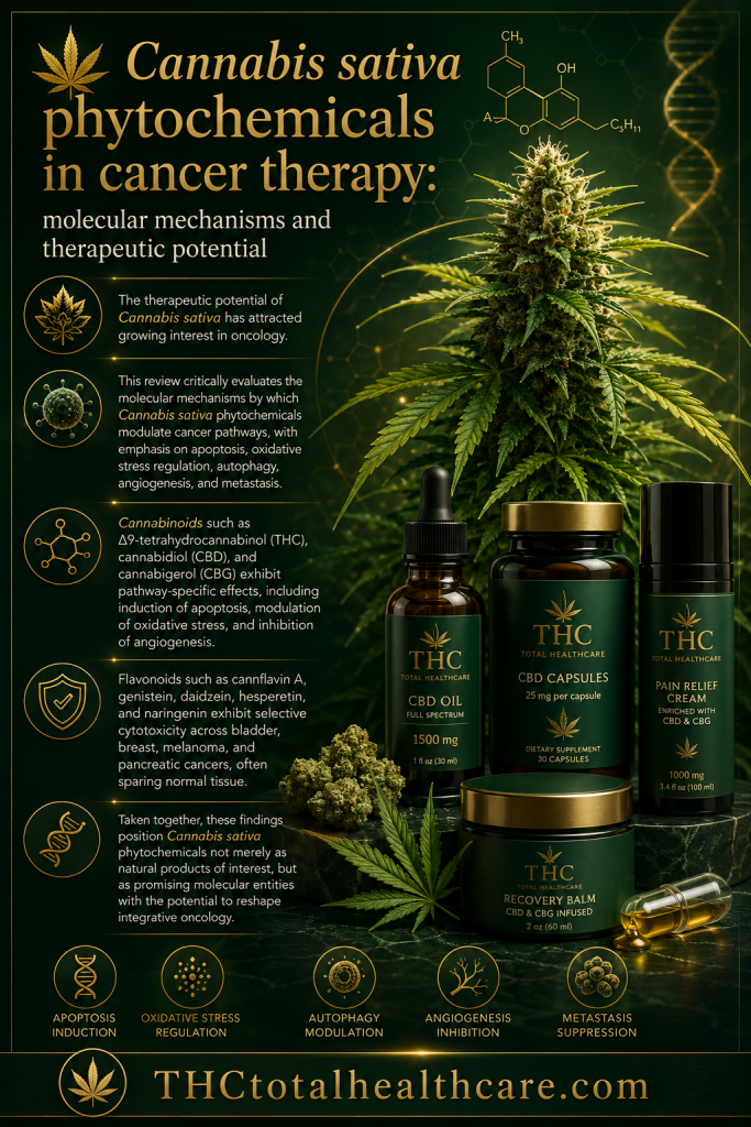
Cannabis cancer research now extends far beyond THC and CBD alone. This 2026 review examines how cannabinoids, flavonoids, and other Cannabis sativa phytochemicals interact with molecular pathways involved in tumor growth, including apoptosis, oxidative stress, autophagy, angiogenesis, and metastasis. The authors highlight evidence involving THC, CBD, CBG, and several flavonoids across cancers including breast, bladder, melanoma, and pancreatic cancer, while also emphasizing that combinations are not always synergistic and that promising laboratory findings have not consistently translated into clinical success. The review concludes that cannabis-derived compounds remain important candidates for integrative oncology, but their potential will depend on better dosing strategies, rational combinations, targeted delivery, and rigorous clinical evaluation.
“Background: The therapeutic potential of Cannabis sativa has attracted growing interest in oncology. Its diverse phytochemicals, including cannabinoids, flavonoids, and terpenes, interact with oncogenic signaling pathways and the endocannabinoid system influencing tumour progression and therapeutic responses.
Objective: This review critically evaluates the molecular mechanisms by which Cannabis sativa phytochemicals modulate cancer pathways, with emphasis on apoptosis, oxidative stress regulation, autophagy, angiogenesis, and metastasis. It also explores synergistic and additive interactions among cannabinoids and flavonoids, highlighting their translational relevance.
Key findings: Cannabinoids such as Δ9-tetrahydrocannabinol (THC), cannabidiol (CBD), and cannabigerol (CBG) exhibit pathway-specific effects, including induction of apoptosis, modulation of oxidative stress, and inhibition of angiogenesis. Flavonoids such as cannflavin A, genistein, daidzein, hesperetin, and naringenin exhibit selective cytotoxicity across bladder, breast, melanoma, and pancreatic cancers, often sparing normal tissue. Importantly, phytochemical interactions are not uniformly synergistic; while combinations such as THC and CBD amplify apoptotic signaling, others act additively or antagonistically. Clinical formulations such as Nabiximols provide translational evidence of cannabinoid synergy, although outcomes remain context-dependent.
Conclusion: The disconnect between preclinical efficacy and clinical outcomes underscores critical gaps in dosing strategies, patient selection, and combination regimens. Future research should prioritize mechanistic studies, rational phytochemical combinations, and innovative drug delivery systems. Taken together, Cannabis sativa phytochemicals emerge as promising molecular entities with the potential to reshape integrative oncology, provided their therapeutic promise is matched with rigorous, evidence-based evaluation.”
“Taken together, these findings position Cannabis sativa phytochemicals not merely as natural products of interest, but as promising molecular entities with the potential to reshape integrative oncology.”
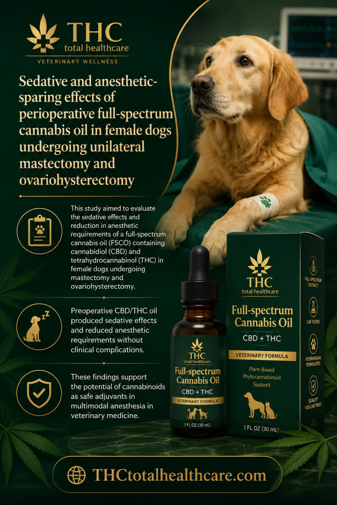
Cannabis-based products are increasingly being studied for their potential to reduce the amount of sedative and anesthetic medication needed during surgery. In this veterinary study, female dogs undergoing mastectomy and ovariohysterectomy received full-spectrum cannabis oil as part of their perioperative care. The researchers found evidence of sedative and anesthetic-sparing effects, suggesting that cannabis oil may influence how much conventional anesthesia is required while also contributing to perioperative pain management. The findings are preliminary and specific to a veterinary surgical setting, but they add to emerging research on cannabinoids as adjuncts to anesthesia and postoperative care.
“The anesthetic management of female dogs with mammary neoplasia, usually classified as ASA II and undergoing invasive procedures such as mastectomy and ovariohysterectomy, requires effective sedation and anesthetic stability due to the increased anesthetic risk associated with advanced age and underlying disease.
In this context, this study aimed to evaluate the sedative effects and reduction in anesthetic requirements of a full-spectrum cannabis oil (FSCO) containing cannabidiol (CBD) and tetrahydrocannabinol (THC) in female dogs undergoing mastectomy and ovariohysterectomy.
Twenty dogs were randomly assigned to two groups: group A (n = 10), treated with FSCO (0.02 mL/kg PO; 0.2 mg/kg CBD and 0.12 mg/kg THC) twice daily for seven days, plus 0.2 mL/kg (2 mg/kg CBD; 1.2 mg/kg THC) one hour before premedication; and group B (n = 10), treated with placebo. Groups A and B had similar ages (9.6; 10.2 years) and weights (7.4; 6.8 kg). Anesthesia was induced with propofol and maintained with sevoflurane. Outcomes included sedation scores, anesthetic requirements, rescue analgesia, responses to instrumentation, and adverse effects. The treated group required less propofol (2.33 vs. 5.98 mg/kg; p = 0.001) and lower sevoflurane concentrations from T0 to T4 (p < 0.05). Sedation scores were higher at 40 and 60 min (median of 4 vs. 0, and 6.5 vs. 0.5; p = 0.015 and p = 0.002, respectively). Fewer treated dogs required rescue analgesia (3/10 vs. 6/10; p = 0.178). No differences were observed in catheterization, intubation, or adverse effects.
Preoperative CBD/THC oil produced sedative effects and reduced anesthetic requirements without clinical complications.
These findings support the potential of cannabinoids as safe adjuvants in multimodal anesthesia in veterinary medicine.”
“The findings of this study indicate that preoperative administration of full-spectrum Cannabis sativa oil containing CBD and THC exerts significant sedative and anesthetic-sparing effects in female dogs with mammary neoplasia undergoing mastectomy and ovariohysterectomy.
The use of this phytocannabinoid extract significantly reduced the required doses of both propofol for induction and sevoflurane for maintenance, supporting its potential as an adjuvant in multimodal anesthetic protocols. Furthermore, the therapy was not associated with clinical adverse effects or compromised anesthetic safety, suggesting a favorable safety profile for perioperative use in oncological patients.”
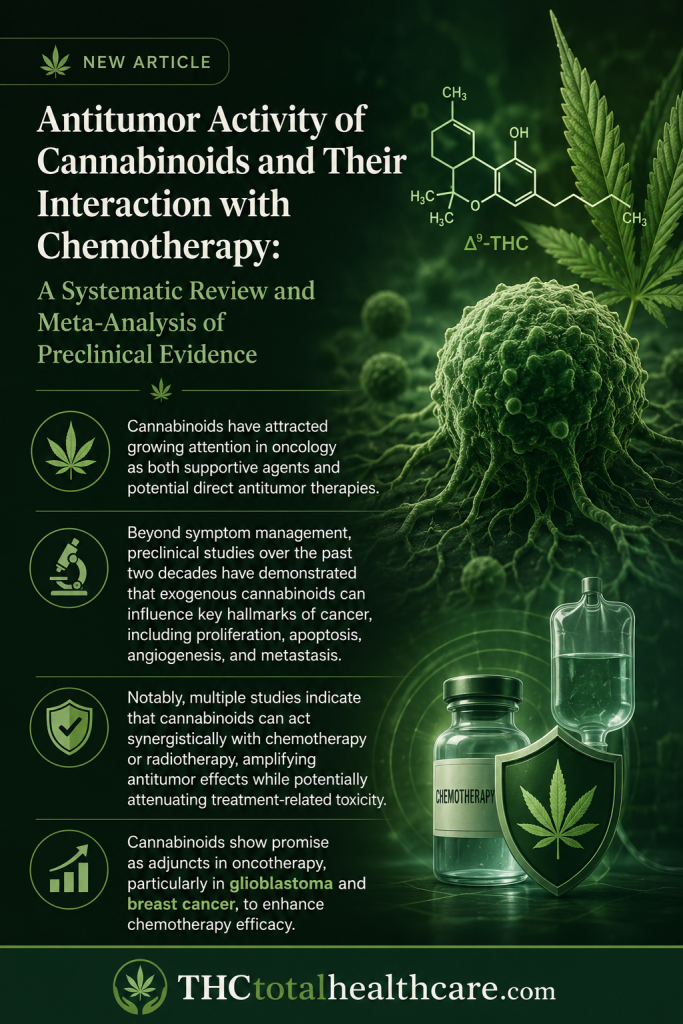
Cannabinoids are increasingly being studied for direct antitumor effects and for their ability to influence the response to conventional chemotherapy. This systematic review and meta-analysis of preclinical research found that cannabinoids significantly reduced tumor growth and cancer-cell viability across multiple models. The analysis also identified evidence that cannabinoids can enhance the effectiveness of several chemotherapy agents, supporting the possibility of useful combination strategies. The findings strengthen the case for cannabinoids as biologically active anticancer compounds with potential both as direct therapies and as adjuncts to standard treatment.
“Background: Cannabinoids are studied as anticancer agents, but their effects vary across tumors, compounds, and experimental settings, underscoring the need to define consistent patterns. Our objective was to map cannabinoid efficacy across cancer preclinical models and identify tumor settings with the greatest translational promise.
Methods: The protocol was registered on PROSPERO (CRD42025543744); PubMed, Embase, and CENTRAL were searched on 4 April 2024 for in vitro and in vivo studies assessing cannabinoid antitumor effects alone or with chemotherapy versus vehicle or chemotherapy only. Random-effects models yielded pooled mean differences (MD) with 95% confidence intervals (CI). MDs of viable cells were calculated for in vitro assays and tumor volume (mm3) for in vivo studies. Reports of various compounds, cannabidiol (CBD), tetrahydrocannabinol (THC) or synthetic cannabinoids, were pooled.
Results: We included 189 studies in the final analysis. In vitro, cannabinoids reduced cell viability modestly overall, with significant effects in glioblastoma (MD -18.77 [CI: -27.15; -10.39]) and a nonsignificant trend in breast cancer (MD -6.75 [CI: -13.90; 0.40]). For in vivo, monotherapy showed the most consistent efficacy in glioblastoma, significantly reducing tumor volume by MD -980.58 mm3; [CI: -1270.2; -690.88]. Addition to temozolomide produced a favorable but nonsignificant decrease of MD -220.65 mm3; [CI: -579.34; 138.03, vs. temozolomide]. In breast cancer, cannabinoids achieved smaller yet significant tumor reductions (MD -402.64 mm3); [CI: -671.84; -133.45]. Synthetic agents had the largest effect (MD -1295.19 mm3); [CI: -1664.33; -928.05] -CBD plus doxorubicin vs. doxorubicin). Lung cancer (MD -562.17 mm3); [CI: -693.99; -430.35] and prostate cancer (MD -1136.59 mm3); [95% CI: -1320.97; -952.21] also had a significant response, whereas colon, pancreatic, and hepatocellular carcinoma models showed inconsistent or null responses.
Conclusions: Cannabinoids show promise as adjuncts in oncotherapy, particularly in glioblastoma and breast cancer, to enhance chemotherapy efficacy. These findings should be interpreted with caution given the high inter-study heterogeneity typical of preclinical research and should be considered hypothesis-generating, warranting further validation in standardized and clinically relevant models.”
“Cannabinoids have attracted growing attention in oncology as both supportive agents and potential direct antitumor therapies.”
“Beyond symptom management, preclinical studies over the past two decades have demonstrated that exogenous cannabinoids can influence key hallmarks of cancer, including proliferation, apoptosis, angiogenesis, and metastasis.”
“Notably, multiple studies indicate that cannabinoids can act synergistically with chemotherapy or radiotherapy, amplifying antitumor effects while potentially attenuating treatment-related toxicity. These interactions are clinically appealing, as they suggest a capacity to sensitize tumor cells to conventional agents and possibly enable dose reductions that limit systemic adverse effects.”
“Cannabinoids show emerging potential as adjuncts in oncological treatment, with relatively consistent signals observed particularly in glioblastoma and breast cancer models.”