Transient CB2R modulation durably restricts breast cancer plasticity by stabilizing a luminal like cell identity

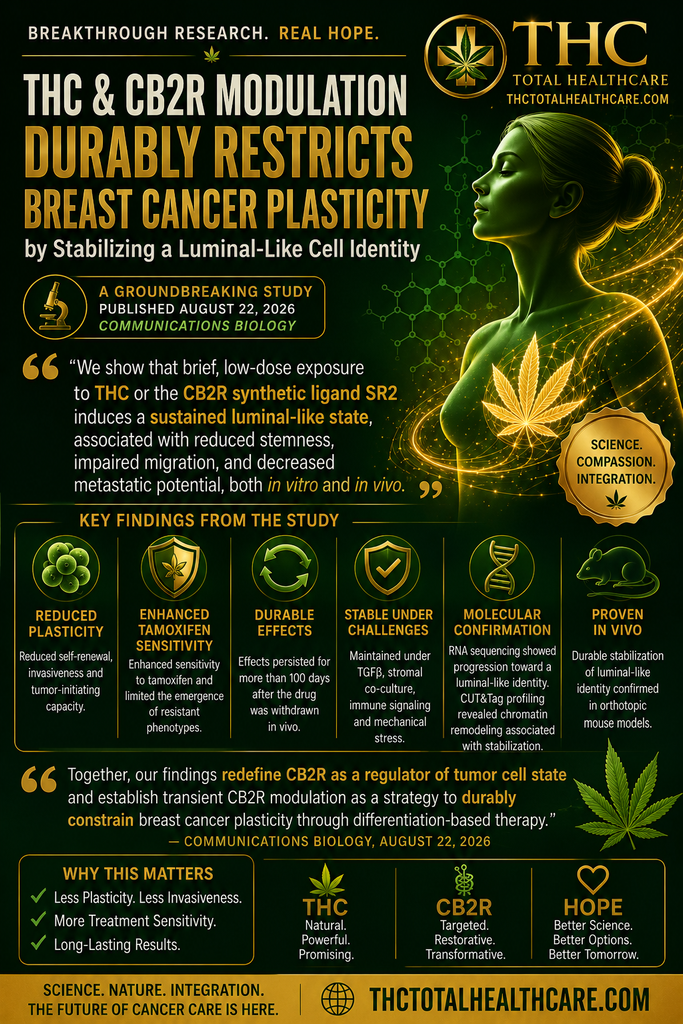
New breast cancer research suggests that brief modulation of cannabinoid receptor 2 (CB2R), including with THC, may have lasting effects on tumor cell behavior. Researchers found reduced self-renewal, invasiveness, and tumor-initiating capacity, along with greater tamoxifen sensitivity and a durable shift toward a more stable luminal-like cell identity.

“Cancer cell plasticity enables therapy resistance and metastasis by allowing transitions between stem-like and differentiated states. Differentiation-based strategies aim to stabilize tumor cell identity, yet pharmacological approaches that induce durable lineage restriction remain limited.

Here, we show that transient modulation of cannabinoid receptor 2 (CB2R) durably restricts breast cancer cell plasticity by stabilizing a luminal-like cell state. Using patient-derived and murine tumor organoids, we demonstrate that brief, low-dose CB2R modulation reduces self-renewal, invasiveness and tumor-initiating capacity, while enhancing tamoxifen sensitivity and limiting the emergence of resistant phenotypes.

These effects persist under pro-dedifferentiation conditions, including TGFβ exposure, stromal co-culture, immune signaling and mechanical stress, and are maintained in vivo following orthotopic transplantation. RNA sequencing reveals a progressive transition from an early plastic state toward a stabilized luminal-like identity, supported by CUT&Tag profiling that uncovers chromatin remodeling associated with this stabilization.

Together, our findings redefine CB2R as a regulator of tumor cell state and establish transient CB2R modulation as a strategy to durably constrain breast cancer plasticity through differentiation-based therapy.”

https://www.nature.com/articles/s42003-026-10837-1

“Low-dose THC keeps breast cancer cells in tumor models from reverting to an aggressive state”

https://medicalxpress.com/news/2026-09-dose-thc-breast-cancer-cells.html

Brain cannabinoid CB1 receptor signaling modulates reward responses and inhibitory control in humans

Researchers using PET and fMRI brain imaging found that natural differences in cannabinoid CB1 receptor availability are associated with how strongly the brain responds to anticipated food rewards and how it engages inhibitory-control systems. The findings provide new human evidence connecting the endocannabinoid system with appetite-related behavior and suggest CB1 signaling as a potential therapeutic target for eating disorders and appetite regulation.

“The central endocannabinoid system, particularly the in vivo cannabinoid type 1 (CB1) receptor signaling, presents a promising target for treating eating disorders. However, its precise role in appetite control remains unclear.

This study aimed to determine how CB1 receptor signaling contributes to key aspects of appetite regulation, specifically anticipatory food reward responses and inhibitory control.

Forty-one healthy male participants underwent [18F]FMPEP-d2 positron emission tomography (PET) to quantify CB1 receptor availability. Functional magnetic resonance imaging (fMRI) was used to assess anticipatory neural responses to food cues, while inhibitory control was measured using a go/nogo task.

Data show that individuals with higher CB1 receptor availability exhibited stronger anticipatory reward-related neural responses and reduced activation during inhibitory control. Therefore, CB1 receptor signaling plays a distinct role in modulating reward and inhibitory processes related to feeding behavior.

The findings suggest that the CB1 receptor may serve as a therapeutic target for regulating appetite.”

https://pubmed.ncbi.nlm.nih.gov/42662580

“Brain cannabinoid CB1 receptor availability modulates reward responses and inhibitory control, key neural mechanisms involved in appetite regulation. The findings suggest that individuals with elevated CB1 receptor availability may require increased engagement of inhibitory control mechanisms to counteract reward-driven responses triggered by external cues, as opposed to enhanced internal satiety signals.

These insights have potential implications for developing therapeutic strategies targeting the endogenous cannabinoid system to address obesity epidemic.”

https://direct.mit.edu/imag/article/doi/10.1162/IMAG.a.1344/138067/Brain-cannabinoid-CB1-receptor-signaling-modulates

Differential Effects of Cannabidiol and Cannabigerol on Cognition, Neuroinflammation, and Blood-Brain Barrier Integrity in a Rat Model of Iron Overload

Researchers comparing cannabidiol (CBD) and cannabigerol (CBG) found that both cannabinoids reversed recognition-memory impairment and restored a protein important to blood-brain barrier integrity in rats exposed to excessive iron early in life.

Both CBD and CBG reduced the inflammatory marker IL-1β, although their broader effects on inflammation differed. The findings suggest that the two phytocannabinoids may protect cognitive function through distinct but complementary mechanisms involving neuroinflammation and the blood-brain barrier.

“Iron is an essential micronutrient for brain development, participating in mitochondrial respiration, myelination, and neurotransmitter synthesis. However, previous studies have demonstrated that excessive iron during early postnatal life induces oxidative reactions, leading to mitochondrial dysfunction and synaptic failure. These alterations compromise energy metabolism and neuronal integrity, contributing to long-lasting cognitive dysfunction and increased brain vulnerability later in life.

This study evaluated the effects of cannabidiol (CBD) and cannabigerol (CBG) on behavioral, neuroinflammatory, and blood-brain barrier (BBB) outcomes in rats exposed to early-life iron overload.

Male Wistar rats received iron carbonyl (30 mg/kg, intragastrically) from postnatal day 12 to 14. At three months of age, they were treated intraperitoneally with CBD, CBG (both at 10 mg/kg), or vehicle for 21 days. Cognitive performance was assessed in the open field and object recognition tasks. We examined hippocampal levels of interleukin-1 beta (IL-1β), interleukin-6 (IL-6), tumor necrosis factor alpha (TNF-α), as proinflammatory markers, and occludin, a protein known to regulate BBB permeability.

Iron-exposed animals showed impaired recognition memory, with elevated TNF-α and IL-1β, while CBD reversed memory deficits and reduced IL-1β in iron-treated animals, without affecting TNF-α.

CBG restored memory, decreased IL-1β in both iron-treated and controls, and increased TNF-α in controls. Also, iron overload reduced occludin expression in vehicle-treated rats which was reversed by both CBD and CBG.

These findings highlight inflammation and BBB disruption as mediators of iron-induced cognitive dysfunction and show that both phytocannabinoids act through distinct but complementary mechanisms, supporting their therapeutic potential in neuroinflammation linked to iron overload.”

https://pubmed.ncbi.nlm.nih.gov/42663810

https://link.springer.com/article/10.1007/s12640-026-00826-x


Chronic THC exposure modulates behavioral outcomes and endocannabinoid signaling in HIV-1 Tg26 mice in a sex-dependent manner

Researchers examined how chronic THC exposure affects behavior and endocannabinoid signaling in an HIV-1 mouse model.

Chronic THC helped attenuate the decline in motor coordination and was associated with increased CB1 receptor expression in the cerebellum. The study also found major sex-dependent differences, with female and male mice showing different patterns of motor impairment and endocannabinoid signaling.

THC did not produce detectable pain-relieving effects in this model, but the findings suggest that chronic THC can influence HIV-related neurological changes through the endocannabinoid system.

Overall, the study highlights the complex relationship between THC, HIV-associated neurological dysfunction, and biological sex.

“While combined antiretroviral therapy (cART) has transitioned HIV-1 into a manageable chronic condition, it fails to eradicate latent viral reservoirs in the central nervous system (CNS) that drive persistent neuroinflammation and synaptodendritic injury. Consequently, people living with human immunodeficiency virus type-1 (HIV-1) often utilize cannabis to manage neurological symptoms, yet the long-term impact of exogenous cannabinoids on the HIV-1-burdened brain remains poorly understood.

In this study, we utilized the HIV-1 Tg26 mouse model to evaluate how chronic Δ9-tetrahydrocannabinol (THC, 3mg/kg) exposure influences motor coordination, thermal nociception, and endocannabinoid (eCB) signaling in the context of constitutive viral protein expression.

Our results demonstrate that HIV-1 viral protein expression was associated with impaired acquisition of cerebellum-dependent motor learning in a sex-dependent manner. This deficit was primarily driven by females and coincided with altered markers of eCB plasticity, characterized by elevated monoacylglycerol lipase (MAGL) expression and a depletion of 2-arachidonoylglycerol (2-AG). Conversely, males exhibit increased cerebellar CB1R and CB2R expression, which paralleled preserved rotarod performance. In the spinal cord, viral protein expression was associated with thermal hyposensitivity and a reduction in 2-AG and cannabinoid receptor levels, a pattern consistent with HIV-1-associated alterations in sensory processing circuits.

While chronic THC failed to produce detectable antinociceptive effects, consistent with spinal CB1R downregulation, it successfully attenuated the temporal decline of motor coordination with upregulating cerebellar CB1R. Data from a separate acute THC cohort demonstrated detectable THC and metabolite concentrations in plasma and cortex, while also revealing sex- and genotype-dependent differences in these measures.

Together, these findings identify sex-specific eCB signaling as a critical factor associated with the neurobiological response to HIV-1 proteins and provide a biological framework for understanding sex-dependent variability in cannabinoid efficacy.”

https://pubmed.ncbi.nlm.nih.gov/42660238

“Collectively, these results provide a biological framework for understanding sex-dependent variability in cannabinoid responses and support the inclusion of sex as a key factor in the development of cannabinoid-based adjunct therapies for chronic neuroinflammatory conditions.”

https://www.sciencedirect.com/science/article/pii/S0361923026003862?via%3Dihub

The Potential of Cannabidiol for the Treatment of Psychosis: Endocannabinoid Regulation of Serotonergic Neurotransmission via Serotonin 5-HT1A/2A Receptors

Psychosis involves complex changes in brain signaling, including systems regulated by serotonin and the endocannabinoid system. Cannabidiol (CBD) has drawn growing interest because it may influence both.

This review examines how CBD may affect serotonergic neurotransmission through 5-HT1A and 5-HT2A receptors, while also interacting with the endocannabinoid system in ways relevant to psychotic disorders.

The evidence supports continued investigation of CBD as a potential therapeutic approach for psychosis and related symptoms.

“Cannabis, widely used for both medical and non-medical purposes, primarily contains Δ9-tetrahydrocannabinol (Δ9-THC) and cannabidiol (CBD).

This review aims to clarify how the molecular and receptor-level mechanisms of CBD, compared with Δ9-THC, shape its distinct therapeutic profile and low liability for addiction, thereby informing the safer clinical application of CBD-containing interventions to schizophrenia.

CBD exerts sedative and therapeutic effects, including in treatment-resistant epilepsy, and acts in part via CB1 and CB2 receptors that also mediate Δ9-THC actions. While Δ9-THC activates these receptors as an agonist, triggering Gi-coupled signaling cascades such as MAPK pathways and suppressing presynaptic monoamine release, CBD functions as a non-competitive negative allosteric CB1 modulator and non-competitive antagonist of Δ9-THC. These distinct intracellular signaling mechanisms are considered to underlie the markedly different pharmacological profiles and abuse liabilities of Δ9-THC and CBD.

CBD also engages multiple CNS targets beyond CB1 and CB2, including GPR55, TRPV1 and ENT1, which are implicated in anticonvulsant, anti-inflammatory, analgesic and neuroprotective actions. Although GPR55 pharmacology remains controversial and Δ9-THC shows inconsistent activity at this receptor, converging evidence indicates that CBD can antagonize CB1/CB2 and modulate atypical cannabinoid-sensitive proteins. Excessive activation of certain serotonin receptors, including 5-HT1A/2A, are thought to underlie some symptoms of psychosis.

These diverse molecular actions are thought to underlie CBD’s broad therapeutic potential across seizures and other CNS disorders, providing a mechanistic rationale for exploring CBD as a treatment option for psychosis in schizophrenia and stimulant-induced psychotic states.”

https://pubmed.ncbi.nlm.nih.gov/42658346/

https://link.springer.com/article/10.1007/s11064-026-04860-1

Multiple binding modes underlie Cannabis sativa cannabinoids recognition by peroxisome proliferator-activated receptor gamma

Cannabinoids interact with more than the classical cannabinoid receptors CB1 and CB2. One important target is PPARγ, a nuclear receptor involved in metabolism, inflammation, insulin sensitivity and several disease-related pathways.

In this study, researchers examined how multiple Cannabis sativa cannabinoids bind to and activate PPARγ. The results showed that different cannabinoids can engage the receptor through multiple binding modes, with acidic cannabinoids such as THCA and CBDA showing particularly strong activity.

The findings help clarify another molecular pathway through which cannabis compounds may influence biological processes relevant to metabolic, inflammatory and other disorders.

Introduction: Peroxisome proliferator-activated receptor gamma (PPARγ) is a ligand-activated nuclear receptor with broad therapeutic relevance across various pathologies, including type 2 diabetes, obesity, cancer, and inflammatory disorders. Cannabinoids are a class of terpene-phenolic compounds from Cannabis sativa L. that have been shown to act as partial agonists of PPARγ. Among them, the acidic forms Δ9-tetrahydrocannabinolic acid (THCA) and cannabidiolic acid (CBDA) display higher potency than their decarboxylated counterp arts Δ9-tetrahydrocannabinol (THC) and cannabidiol (CBD). Despite experimental evidence supporting direct PPARγ-cannabinoid interaction, the molecular determinants governing ligand recognition within the binding pocket have not yet been comprehensively investigated.

Methods: A combination of molecular docking and molecular dynamics simulations was employed to characterize the binding modes of THC, CBD, THCA, and CBDA within the PPARγ ligand-binding domain. Docking calculations were performed on a curated set of 70 PPARγ crystal structures co-crystallized with structurally diverse ligands, exploiting thus the conformational variability of the binding pocket. The best-ranked solutions were subjected to 500 ns MD simulations and evaluated on the basis of ligand stability, persistence of polar and aromatic-aromatic interactions with the receptor, and energetic contributions estimated by MM/GBSA. Three candidate binding modes per ligand were selected and their trajectories extended to 1,000 ns.

Results: All four cannabinoids yielded at least one stable binding mode at the microsecond timescale. The cannabinoids THCA and CBDA displayed a greater number of stable binding modes than THC and CBD, a result consistent with the higher potency previously reported for these compounds in experimental studies. This behavior may be attributable to the formation of salt bridges with basic residues in the binding pocket.

Conclusion: Our findings provide a structural framework for understanding cannabinoid recognition by PPARγ. The ability of these compounds to adopt multiple binding modes may contribute to their partial agonist profile, opening new avenues for the rational design of selective PPARγ modulators with improved therapeutic properties.”

https://pubmed.ncbi.nlm.nih.gov/42639023

“This work provides a foundation for future studies addressing the molecular basis of cannabinoid action on PPARγ.

Moreover, these findings could serve as a starting point to explore the allosteric mechanism by which these small lipophilic molecules influence receptor structure and dynamics.

Finally, the strategy described here may also be applied to determine the binding modes of these natural compounds in other members of the nuclear receptor family, including PPARα and PPARδ, as well as to investigate the binding modes of minor cannabinoids, such as cannabichromene and cannabigerol.”

https://www.frontiersin.org/journals/bioinformatics/articles/10.3389/fbinf.2026.1893303/full


Cannabinoid neuroprotection and cardiotoxicity in retinal disease: the role of the THC:CBD ratio

Cannabinoids are increasingly being studied for their potential to protect retinal cells from damage, but their effects may depend heavily on the balance between THC and CBD.

This study examined different THC-to-CBD ratios in models of retinal disease, assessing both neuroprotective effects in the eye and potential cardiovascular toxicity. The results suggest that cannabinoid composition can significantly influence both therapeutic benefit and safety.

The findings highlight the importance of studying specific THC/CBD ratios rather than treating all cannabinoid formulations as biologically equivalent.

“Cannabinoids, compounds acting on the endocannabinoid system (ECS) and the broader endocannabinoidome, have demonstrated promising neuroprotective effects in retinal neurodegenerative diseases including glaucoma and age-related macular degeneration (AMD).

This review evaluates the evidence for cannabinoid receptor expression and function in the retina, the capacity of cannabinoids to reduce oxidative stress and neuroinflammation, and outcomes in preclinical disease models.

Cannabidiol (CBD) and Δ9-tetrahydrocannabinol (THC) act through cannabinoid receptors 1 and 2 (CB1 and CB2) receptors, as well as non-canonical pathways including transient receptor potential cation channel subfamily V member 1 (TRPV1), G-protein coupled receptor 3 (GPR3), peroxisome proliferator-activated receptor gamma (PPARγ), to confer neuroprotective benefits.

Critically, previously identified neuroprotective effects of cannabis may be substantially masked or reversed by the dramatically rising THC:CBD ratio in commercial strains, which has increased from approximately 10:1 in 2000 to 100:1 in many contemporary products.

This shift means that modern cannabis is biochemically distinct from the preparations used in foundational neuroprotection studies.

CBD-dominant formulations, or preparations maintaining lower THC:CBD ratios, represent a valid therapeutic direction for acute retinal neuroprotection, while the proven safety and efficacy of vitamins such as Age-Related Eye Disease Study 2 (AREDS2) therapy provide a reliable chronic neuroprotective strategy.

While the biological plausibility and animal evidence for CBD/THC protection is good, human trials are needed to validate any use in retinal diseases.”

https://www.academia.edu/3071-4087/2/3/10.20935/AcadNeurosci8479


Beyond inflammation: Cannabinoid receptors as metabolic checkpoints in glial reprogramming during neurodegeneration

Cannabinoid receptors are increasingly being studied for roles that go beyond controlling inflammation in the brain.

This review examines how CB1 and CB2 receptors may act as metabolic checkpoints in glial cells, influencing how these cells use energy, respond to stress and change their behavior during neurodegenerative disease.

The findings highlight a broader role for the endocannabinoid system in shaping brain metabolism and glial function, with potential relevance to conditions such as Alzheimer’s, Parkinson’s and other neurodegenerative disorders.

“Cannabinoid receptors have traditionally been regarded as regulators of neuroinflammation. However, their anti-inflammatory effects alone are insufficient to fully elucidate their complicated roles in neurodegenerative diseases (NDDs).

Mounting evidence identifies disruptions in energy metabolism as key drivers of neurodegeneration, which has prompted a re-evaluation of the cannabinoid receptor system within the context of brain energy metabolism and pathophysiological processes. Accumulated findings from several independent preclinical studies have offered novel insights into the potential involvement of cannabinoid receptors in energy metabolism, mitochondrial function, and glial metabolic reprogramming.

This review focuses on the metabolic regulatory potential of classical cannabinoid receptors, including cannabinoid receptor type 1 (CB1R) and cannabinoid receptor type 2 (CB2R), with particular attention to mitochondrial CB1 receptors (mtCB1), as well as non-classical targets such as G protein-coupled receptor 55 (GPR55), G protein-coupled receptor 119 (GPR119), and peroxisome proliferator-activated receptors (PPARs). Additionally, the review explores the mechanisms by which astrocytes maintain neuronal support through cell-type-specific metabolic specialization and how metabolic reprogramming in microglia modulates neuroinflammatory phenotypes.

Although the current evidence is predominantly derived from preclinical models and several mechanistic links remain to be experimentally validated, we propose a novel conceptual model, namely the cannabinoid receptor-glial metabolic reprogramming-neuronal metabolic support failure axis, in which the breakdown of metabolic checkpoints is viewed as a crucial event in disease progression.

At the therapeutic level, we advocate shifting from conventional anti-inflammatory approaches to metabolic repair strategies, while also exploring emerging directions in the development of cannabinoid-based medications targeting metabolism.

Collectively, a deep understanding of the complex metabolic regulatory functions of cannabinoid receptors is critical for developing next-generation treatment strategies for NDDs.”

https://pubmed.ncbi.nlm.nih.gov/42624021

“Shift from anti-inflammation to metabolic repair for cannabinoid NDD therapy.”

https://www.sciencedirect.com/science/article/abs/pii/S1567576926011458?via%3Dihub


Endocannabinoid system attenuates emotional stress-induced orofacial musculoskeletal pain in rats

Emotional stress can intensify orofacial musculoskeletal pain, but the endocannabinoid system may help regulate that response. In this rat study, researchers found that activating cannabinoid signaling reduced stress-related facial pain, supporting a role for the endocannabinoid system in controlling the interaction between emotional stress and pain.

“Orofacial musculoskeletal pain (OMP) is a common feature of temporomandibular disorders (TMD), a group of conditions affecting the temporomandibular joint, chewing muscles, and associated structures.

The etiology of muscular TMD is currently understood within a biopsychosocial model of pain, highlighting the complexity related to OMP. In this context, the objective of this study was to investigate the OMP induced by psychological/emotional stress (ES) in rats, regarding the role of the endocannabinoid system (ECS) through CB1 and CB2 receptors.

Male Wistar rats were divided into Control and ES groups. OMP was induced by ES using the communication box model and evaluated through the mechanical threshold in masticatory muscles. c-Fos, CB1, and CB2 immunostaining was evaluated in the trigeminal ganglion (TG) and in the trigeminal nucleus caudalis (Sp5C). The selective CB1 or CB2 antagonist (AM251 or AM630, respectively) or the cannabinoid receptor non-selective agonist (WIN55,212-2) were administered to both groups, and the OMP was evaluated. The mechanical sensitivity of the masticatory muscles increased in the ES group, accompanied by increased c-Fos expression in the TG and Sp5C. AM251 and AM630 increased mechanical sensitivity, while WIN55,212-2 decreased the OMP. Furthermore, ES increased CB1 density in the TG and elevated both CB1 and CB2 in the Sp5C. Additionally, CB2 was increased in the masseter muscle.

Thus, cannabinoid receptors played a protector role in OMP caused by ES, indicating that cannabinoid drugs or the modulation of the ECS may represent a promising approach for the treatment of OMP frequently observed in patients with TMD.”

https://pubmed.ncbi.nlm.nih.gov/42585375

https://www.scielo.br/j/bjmbr/a/DHTkCSCk5bcNGJrbWcb67Jz/?lang=en


Priming Canine Adipose Tissue-Derived Mesenchymal Stem Cells with CBD-Rich Cannabis Extract Modulates Neurotrophic Factors Expression Profile

Cannabidiol-rich cannabis extracts may influence the therapeutic behavior of mesenchymal stem cells before they are used in regenerative treatments. In this laboratory study, priming canine adipose-derived mesenchymal stem cells with a CBD-rich extract altered the expression of neurotrophic factors involved in neuronal survival, growth, and repair, suggesting a potential strategy for enhancing stem-cell-based therapies for neurological injury or disease.

“The endocannabinoid system regulates key biological functions such as neuroprotection, pain modulation, inflammation, and immunomodulation.

Cannabis-based therapies have gained attention due to the therapeutic potential of their bioactive compounds, particularly phytocannabinoids like cannabidiol (CBD), which exhibit anti-inflammatory, neuroprotective, and immunomodulatory properties.

Mesenchymal stem cells (MSCs) are widely studied for their regenerative and immunomodulatory potential.

This study evaluated the effects of priming canine adipose tissue-derived MSCs (cAT-MSCs) with a CBD-rich cannabis extract on cell morphology, viability, neurotrophic factor gene expression, and cytokine gene and protein expression.

cAT-MSCs (n = 5) were primed for 24 h and divided into three groups: Control (C, unprimed), D1 (2.25 µM CBD), and D2 (225 nM CBD). No morphological or viability changes were observed. Gene expression analysis showed that groups D1 and D2 exhibited increased HGF expression. D1 also showed increased IDO and decreased BDNF expression. In contrast, no significant changes were observed in GDNF, IL-10, TNF-α, IFN-γ, or PTGES2. Regarding the cytokine profile, GM-CSF, IL-2, and IL-10 were undetectable. Notably, IL-8 and MCP-1 levels were significantly reduced in D1 compared to the control.

These findings suggest that CBD priming modulates key regenerative and inflammatory mediators in cAT-MSCs, supporting its potential application in enhancing the efficacy of cell-based therapies.”

https://pubmed.ncbi.nlm.nih.gov/41150065

“Mesenchymal stem cells (MSCs) are used in veterinary medicine for their regenerative, immunomodulatory, and anti-inflammatory properties. Compounds from cannabis, especially cannabidiol (CBD), have shown promising anti-inflammatory and healing effects.

This study evaluated whether a CBD-rich cannabis extract modulates important regenerative and inflammatory factors in MSCs derived from canine adipose tissue. After priming canine adipose tissue-derived MSCs for 24 h, we found no changes in their morphology or viability. However, the priming with CBD-rich cannabis extract has increased the activity of certain genes linked to tissue repair and reduced the levels of inflammatory cytokines.

These results suggest that CBD can influence key factors that help stem cells repair tissue and control inflammation, potentially improving their use in future veterinary therapies.”

https://www.mdpi.com/2306-7381/12/10/926