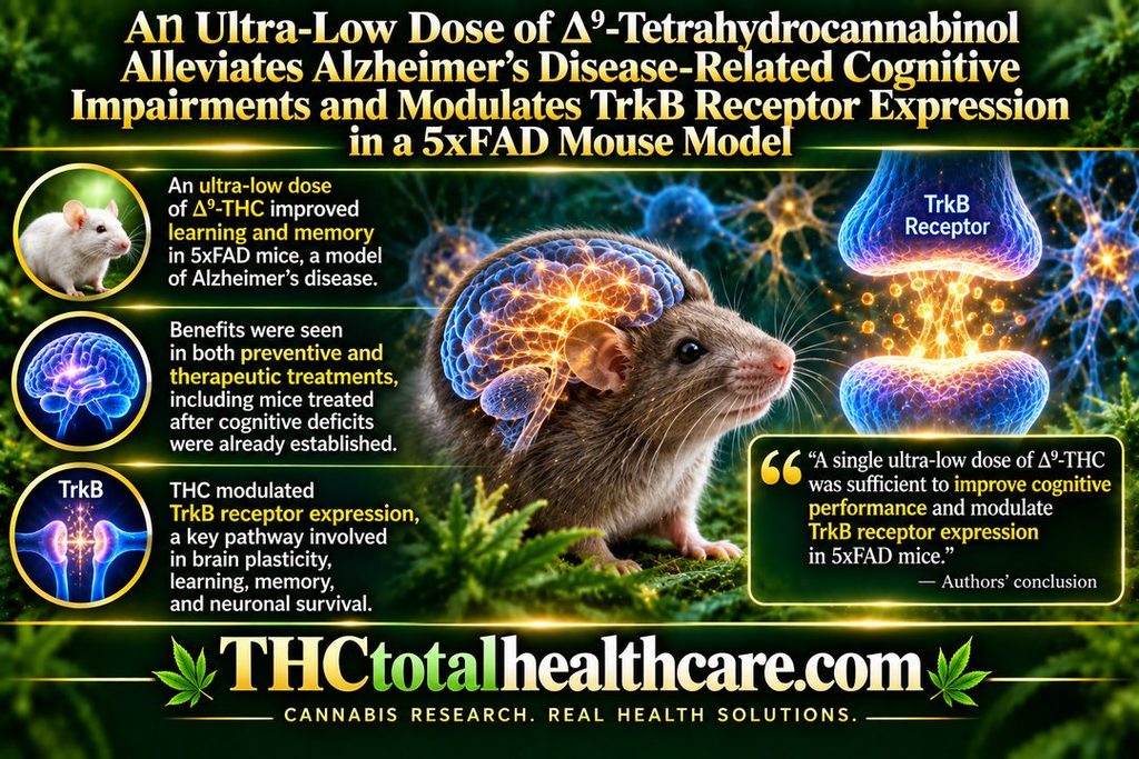
An Ultra-Low Dose of ∆9-Tetrahydrocannabinol Alleviates Alzheimer’s Disease-Related Cognitive Impairments and Modulates TrkB Receptor Expression in a 5XFAD Mouse Model

An ultra-low dose of Δ9-THC improved Alzheimer’s disease-related cognitive impairment in 5xFAD mice, including animals treated after memory problems were already established. The study found improvements in learning and memory without relying on repeated high-dose THC exposure.

The researchers also found changes in TrkB receptor expression, a signaling pathway closely tied to brain plasticity, learning, memory, and neuronal survival. These changes suggest that THC’s effects may involve more than temporary symptom relief and may influence mechanisms connected to cognitive function.

The findings add to a growing body of preclinical research showing that very low doses of THC can produce different biological effects than higher doses, particularly in the aging and Alzheimer’s-disease brain. In this model, a single ultra-low dose was enough to improve cognitive performance and alter a key neurotrophic signaling pathway.

“Alzheimer’s disease (AD) is the most common form of dementia, but there is still no available treatment.

Δ9-tetrahydrocannabinol (THC) is emerging as a promising therapeutic agent.

Using THC in conventional high doses may have deleterious effects. Therefore, we propose to use an ultra-low dose of THC (ULD-THC). We previously published that a single injection of ULD-THC ameliorated cognitive functioning in several models of brain injuries as well as in naturally aging mice.

Here, 5xFAD AD model mice received a single treatment of ULD-THC (0.002 mg/kg) after disease onset and were examined in two separate experiments for cognitive functions, neurotropic, and inflammatory factors in the hippocampus.

We show that a single injection of ULD-THC alleviated cognitive impairments in 6- and 12-month-old 5xFAD mice. On the biochemical level, our results indicate an imbalance between the truncated TrkB receptor isoform and the full receptor, with AD mice showing a greater tendency to express the truncated receptor, and ULD-THC improved this imbalance. We also investigated the expression of three AD-related inflammatory markers and found an ameliorating effect of ULD-THC.

The current research demonstrates for the first time the beneficial effects of a single ultra-low dose of THC in a mouse model of AD after disease onset.”

https://pubmed.ncbi.nlm.nih.gov/36012711

“The current research demonstrates for the first time the beneficial effects of a single ultra-low dose of THC in a mouse model of AD after disease onset. As THC is a cheap, widely available substance already approved for use in other conditions, this research brings us closer to understanding its mechanisms and will possibly lead to new treatments.”

https://www.mdpi.com/1422-0067/23/16/9449/htm

Need this THC study explained? thcXplained.com
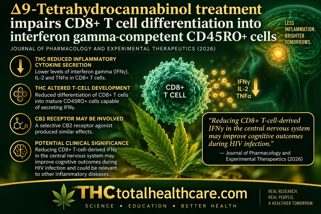
Δ9-Tetrahydrocannabinol treatment impairs CD8+ T cell differentiation into interferon gamma-competent CD45RO+ cells

New research published in The Journal of Pharmacology and Experimental Therapeutics shows that Δ9-tetrahydrocannabinol (THC) can alter the development of CD8+ T cells involved in inflammatory immune responses. Researchers found that THC reduced secretion of interferon gamma (IFNγ), interleukin-2 (IL-2), and tumor necrosis factor alpha (TNFα), three signaling molecules involved in immune activation and inflammation.

Rather than simply preventing cells from producing IFNγ, THC appeared to change how the T cells developed. THC reduced the differentiation of CD8+ T cells into mature CD45RO+ cells capable of secreting IFNγ. A selective CB2 receptor agonist produced similar effects, suggesting that activation of the cannabinoid CB2 receptor may play an important role in THC’s immune-modulating activity.

The researchers examined these effects partly because IFNγ-producing CD8+ T cells contribute to persistent neuroinflammation associated with HIV-associated neurocognitive disorders. They concluded that reducing CD8+ T-cell-derived IFNγ in the central nervous system may potentially improve cognitive outcomes during HIV infection, while noting that the findings may also have relevance to other inflammatory diseases involving IFNγ-producing CD8+ T cells.

“Chronic human immunodeficiency virus (HIV) infection results in a persistent state of neuroinflammation, even with combined antiretroviral therapy. Neuroinflammation contributes to pathology termed HIV-associated neurocognitive disorder, which is exacerbated by interferon gamma (IFNγ)-producing CD8+ T cells in the central nervous system.

Many people living with HIV self-report using Cannabis sativa to mitigate symptoms of chronic infection and side effects of treatment. C. sativa is composed of various phytocannabinoids, including Δ9-tetrahydrocannbinol (THC), which possesses immune-modulating properties.

This study aims to determine whether C. sativa use by people living with HIV, and specifically THC, affects IFNγ secretion by CD8+ T cells.

We found that HIV status does not influence T cell IFNγ responses, and C sativa use modestly reduces the average secretion of IFNγ by CD8+ T cells from HIV+ donors. Treatment of CD8+ T cells with THC and the selective cannabinoid receptor 2 agonist, JWH-015, reduced T-cell cytokine secretion, with THC eliciting greater suppression than cannabidiol.

These results suggest that THC treatment does not directly impair IFNγ gene expression or protein production, as determined by polymerase chain reaction and flow cytometry. However, THC treatment impaired CD8+ T cell differentiation into IFNγ-competent CD45RO+ cells.

These studies are of clinical relevance because reduction of CD8+ T-cell-derived IFNγ in the central nervous system may improve cognitive outcomes during HIV infection. Furthermore, these findings may be generalizable to other inflammatory diseases in which IFNγ-producing CD8+ T cells have been implicated.

SIGNIFICANCE STATEMENT: Δ9-Tetrahydrocannbinol (THC) treatment reduces CD8+ T cell interferon gamma (IFNγ) secretion isolated from HIV- and HIV+ individuals. THC and the selective cannabinoid receptor 2 agonist, JWH-015, both reduced CD8+ T cell secretion of IFNγ, interleukin 2, and tumor necrosis factor α, suggesting the involvement of cannabinoid receptor 2. Although THC treatment does not suppress IFNγ mRNA or protein levels, it reduces the number of IFNγ-secreting cells by impairing CD8+ T-cell differentiation into IFNγ-competent CD45RO+ cells.”

https://pubmed.ncbi.nlm.nih.gov/42748512

https://jpet.aspetjournals.org/article/S0022-3565(26)01210-3/abstract

Need this THC study explained? thcXplained.com

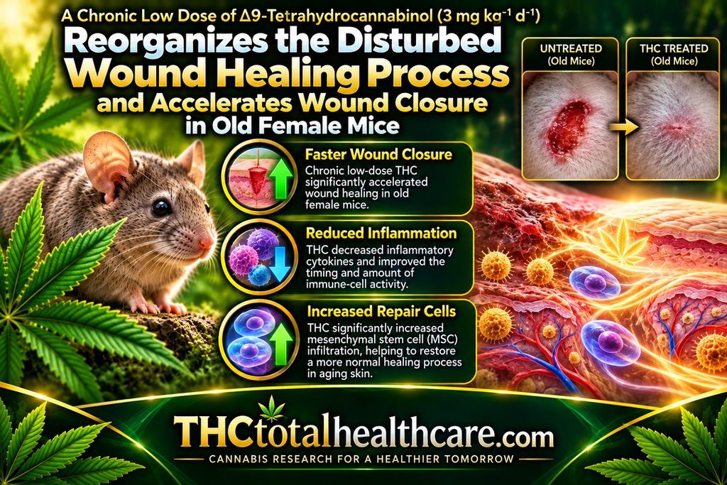
A chronic low dose of Δ9-tetrahydrocannabinol (3 mg / kg / 21 d) reorganizes the disturbed wound healing process and accelerates wound closure in old female mice

A chronic low dose of Δ9-THC accelerated wound closure in old female mice and helped reorganize an age-disrupted healing process. The biggest improvement occurred during the inflammatory phase, when aged wounds normally heal more slowly and remain inflamed longer.

THC changed the timing and amount of immune-cell activity in the wound, reduced inflammatory cytokines, and significantly increased infiltration of mesenchymal stem cells (MSCs) involved in tissue repair. The treatment did not increase growth-factor release, suggesting the benefit came mainly from better coordination of inflammation and cell migration.

The researchers concluded that THC improved wound healing by restoring a more normal sequence of immune-cell infiltration and cytokine release. The findings suggest that stimulating the aging endocannabinoid system may help support skin regeneration and tissue repair in older organisms.

“Wound healing in old mice is characterized by disturbed tissue homeostasis, manifested by delayed immune cell infiltration and reduced growth factor secretion, leading to a delayed onset and prolonged duration of the inflammatory phase.

The endocannabinoid system (ECS) is an important regulator of tissue homeostasis and cell migration and is also considered to be subject to aging processes, which may contribute to observable aging phenomena. Therefore, stimulating the aged ECS could represent a therapeutic option to support tissue regeneration in aging.

Female old mice received a low-dose of medical THC daily for 3 weeks, before four excisional full skin wounds were created. At day 1, 3 and 7 post-surgery, the wound closure rate was analyzed and wound samples were examined immunohistochemically for the numbers of granulocytes, M1-macrophages and mesenchymal stem cells (MSCs). The concentrations of inflammatory cytokines and regenerative growth factors were determined by ELISA.

Administration of THC improved the wound healing rate of old mice between day 1 and 7, which was associated with an altered timing and quantity of infiltrating immune cells and decreased levels of inflammatory cytokines in wound tissue on days 1 and 3 post-injury.

THC treatment significantly increased MSC infiltration but had no effect on the growth factor release.

The present study confirmed the anti-inflammatory activity of THC in vivo.

The THC-treatment improved wound healing in old mice by coordinating the temporal sequence of immune cell infiltration and cytokine release. Thus, restoration of ECS signaling could be an effective strategy to support age-related skin regeneration.”

https://pubmed.ncbi.nlm.nih.gov/40653209/

“A chronic low dose of THC accelerates the wound closure rate during the inflammatory phase in old mice.”

https://www.sciencedirect.com/science/article/pii/S0531556525001615?via%3Dihub

Meed this THC study explained? thcXplained.com

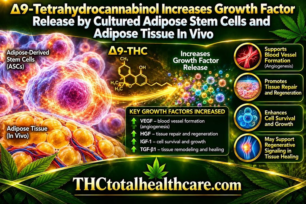
∆9-Tetrahydrocannabinol Increases Growth Factor Release by Cultured Adipose Stem Cells and Adipose Tissue in vivo

Δ9-THC increased the release of growth factors from cultured adipose-derived stem cells and from adipose tissue in vivo. These growth factors are involved in processes such as cell survival, tissue repair, blood-vessel formation, and regeneration.

The researchers found that THC changed the secretory activity of adipose stem cells rather than simply affecting their growth. This suggests that THC may influence how these cells communicate with surrounding tissue and support repair through the factors they release.

The findings point to a potential role for THC in regenerative signaling and tissue-healing biology, particularly through adipose-derived stem cells and the local tissue environment. The study adds another dimension to cannabinoid research by showing that THC can affect not just mature cells, but also the reparative signals produced by stem-cell populations.

“Background: Because of its biocompatibility and its soft and dynamic nature, the grafting of adipose tissue is regarded an ideal technique for soft-tissue repair. The adipose stem cells (ASCs) contribute significantly to the regenerative potential of adipose tissue, because they can differentiate into adipocytes and release growth factors for tissue repair and neovascularization to facilitate tissue survival. The present study tested the effect of administering a chronic low dose of ∆9-tetrahydrocannabinol (THC) on these regenerative properties, in vitro and in vivo.

Methods: Human ASCs were exposed to increasing concentrations of THC. Resazurin conversion was applied to investigate the effect on metabolic activity, cell number was assessed by crystal violet staining, tri-linear differentiation was evaluated by specific colorimetric approaches, and the release of growth factors was analyzed by ELISA. Two groups of mice were treated daily either with a low dose of THC (3 mg/kg) or a vehicle solution. After 3 weeks, adipose tissue was obtained from excised fat deposits, homogenized and tested for growth factor contents.

Results: THC decreased ASC proliferation but increased metabolic activity as well as adipogenic and chondrogenic differentiation. A low concentration of THC (1 µM) enhanced the growth factor release by ASCs. The concentration of these cytokines was also increased in adipose tissue of mice treated with THC.

Conlusion: Our results indicate that chronic activation of the endocannabinoid system promoted differentiation and growth factor release of ASCs, which could be of specific value for enhancing the regenerative potential of adipose tissue.”

https://pubmed.ncbi.nlm.nih.gov/39825991/

“Our findings may offer a new approach for improved application of adipose tissue in regenerative medicine.”

https://link.springer.com/article/10.1007/s13770-024-00692-8

Need this THC study explained? thcXplained.com

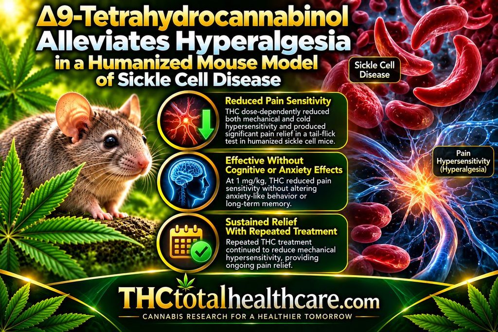
Δ 9 -Tetrahydrocannabinol alleviates hyperalgesia in a humanized mouse model of sickle cell disease

Δ9-THC reduced pain hypersensitivity in a humanized mouse model of sickle cell disease. In mice expressing more than 99% human sickle hemoglobin, acute THC treatment dose-dependently reduced both mechanical and cold hypersensitivity and also produced substantial antinociceptive effects in a tail-flick pain test.

At 1 mg/kg, THC reduced pain sensitivity without altering anxiety-like behavior or long-term memory. With repeated treatment, THC continued to provide sustained relief from mechanical hypersensitivity, although tolerance developed to its effects on cold hypersensitivity.

The researchers concluded that THC may represent a potential option for managing chronic pain associated with sickle cell disease. These findings are especially notable because sickle cell pain is often severe and difficult to control, while current treatment frequently depends on opioid analgesics.

“People with sickle cell disease (SCD) often experience chronic pain as well as unpredictable episodes of acute pain, which significantly affect their quality of life and life expectancy. Current treatment strategies for SCD-associated pain primarily rely on opioid analgesics, which have limited efficacy and cause serious adverse effects.

Cannabis has emerged as a potential alternative, yet its efficacy remains uncertain. In this study, we investigated the antinociceptive effects of Δ9-tetrahydrocannabinol (THC), cannabis’ intoxicating constituent, in male HbSS mice, which express >99% human sickle hemoglobin, and male HbAA mice, which express normal human hemoglobin A, as a control.

Acute THC administration (0.1-3 mg-kg-1, intraperitoneal, i.p.) dose-dependently reduced mechanical and cold hypersensitivity in HbSS, but not HbAA mice. In the tail-flick assay, THC (1 and 3 mg-kg-1, i.p.) produced substantial antinociceptive effects in HbSS mice. By contrast, THC (1 mg-kg-1, i.p.) did not alter anxiety-like behavior (elevated plus maze) or long-term memory (24-h novel object recognition). Subchronic THC treatment (1 and 3 mg-kg-1, i.p.) provided sustained relief of mechanical hypersensitivity but led to tolerance in cold hypersensitivity in HbSS mice.

Together, the findings identify THC as a possible therapeutic option for the management of chronic pain in SCD. Further research is warranted to elucidate its mechanism of action and possible interaction with other cannabis constituents. 

Significance Statement The study explores THC’s efficacy in alleviating pain in sickle cell disease (SCD) using a humanized mouse model. Findings indicate that acute THC administration reduces mechanical and cold hypersensitivity in SCD mice without impacting emotional and cognitive dysfunction. Subchronic THC treatment offers sustained relief of mechanical hypersensitivity but leads to cold hypersensitivity tolerance. These results offer insights into THC’s potential as an alternative pain management option in SCD, highlighting both its benefits and limitations.”

https://pubmed.ncbi.nlm.nih.gov/38955494/

“The results indicate that acute THC administration alleviates mechanical and cold hypersensitivity in HbSS mice in a dose-dependent manner without affecting emotional and cognitive dysfunction also present in these mice.”

https://www.sciencedirect.com/science/article/abs/pii/S0022356524178147

Need this THC study explained? thcXplained.com

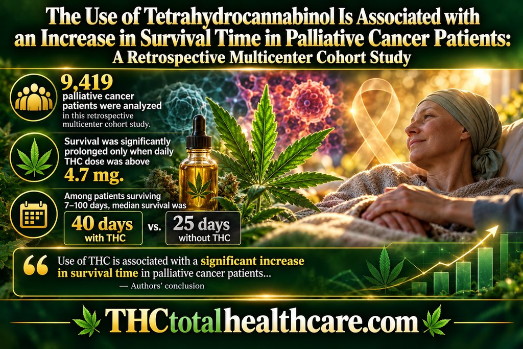
The Use of Tetrahydrocannabinol Is Associated with an Increase in Survival Time in Palliative Cancer Patients: A Retrospective Multicenter Cohort Study

A large retrospective multicenter study of 9,419 palliative cancer patients found that THC use was associated with longer survival. The researchers reported the strongest association among patients receiving more than 4.7 mg of THC per day.

In patients who survived between 7 and 100 days, median survival was 40 days in the THC group compared with 25 days in patients who did not receive THC. The study also examined THC alongside palliative-care treatment rather than as a replacement for standard care.

These findings add an important human-data signal to the growing research on cannabinoids in cancer care. While the study was observational, the association between THC use and longer survival makes this a significant finding for palliative oncology and cannabinoid research.

“Introduction: Tetrahydrocannabinol (THC) is often prescribed for ambulatory palliative patients to improve sleep quality and appetite and to reduce anxiety, stress, and pain. However, it is not known if THC has also an effect on the mortality of these patients.

Method: The objective was the impact of THC on mortality of ambulatory palliative patients. For this purpose, data from the palliative treatment documentation from 5 ambulatory palliative care teams in Brandenburg, Germany were used for this analysis. Survival time was calculated for 3 groups of patients: (1) without THC; (2) with THC in a low dosage (≤4.7 mg per day); and (3) THC in higher doses (≥4.7 mg per day). The analysis was done for 2 cohorts of patients. Cohort 1: all patients with a survival time of at least 7 days after inclusion in specialized ambulatory palliative care (SAPC) and cohort 2: a subgroup of patients with a survival time between 7 and 100 days. Kaplan-Meier curves were created, and multivariate analysis was done to investigate the impact of THC on mortality.

Results: A total of 9,419 patients with a survival time of at least 7 days after inclusion in SAPC were included in the analysis (cohort 1). 7,085 among them had a survival time between 7 and 100 days (cohort 2). In both cohorts, survival time was significantly prolonged by THC, but only when the daily THC dose was above the median of 4.7 mg. Survival time was 15 days longer in cohort 2 (40 vs. 25 days), when more than 4.7 mg THC were prescribed per day.

Conclusion: Use of THC is associated with a significant increase in survival time in ambulatory palliative patients which survive longer than 7 days the initiation of THC prescription and which use of THC >4.7 mg/day.”

https://pubmed.ncbi.nlm.nih.gov/38655402/

“Thus, in view of its significant prolongation of patient survival time, THC therapy should be included as part of the first-line therapy for ambulatory palliative patients.”

https://karger.com/mca/article/7/1/59/896816/The-Use-of-Tetrahydrocannabinol-Is-Associated-with

Need this THC study explained? thcXplained.com

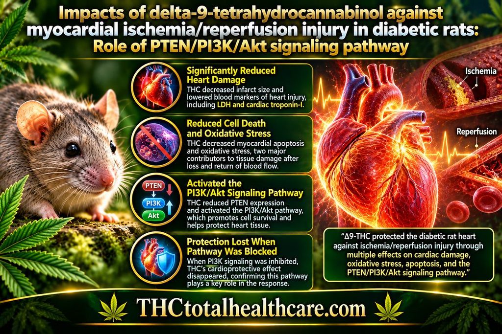
Impacts of delta 9-tetrahydrocannabinol against myocardial ischemia/reperfusion injury in diabetic rats: Role of PTEN/PI3K/Akt signaling pathway

In diabetic rats with myocardial ischemia/reperfusion injury, Δ9-THC significantly reduced heart damage and improved cardiac function. THC pretreatment decreased infarct size as well as blood markers of heart injury, including LDH and cardiac troponin-I.

THC also reduced myocardial apoptosis and oxidative stress, two major contributors to tissue damage when blood flow returns to the heart after a period of ischemia. These protective effects were accompanied by reduced PTEN expression and activation of the PI3K/Akt signaling pathway, which is involved in cell survival.

When researchers blocked PI3K signaling, THC’s cardioprotective effect disappeared, strengthening the evidence that this pathway played an important role in the response. The findings show that THC protected the diabetic rat heart against ischemia/reperfusion injury through multiple measurable effects on cardiac damage, oxidative stress, cell death, and survival signaling.

“Despite the current optimal therapy, patients with myocardial ischemia/reperfusion (IR) injury still experience a high mortality rate, especially when diabetes mellitus is present as a comorbidity. Investigating potential treatments aimed at improving the outcomes of myocardial IR injury in diabetic patients is necessary.

Our objective was to ascertain the cardioprotective effect of delta 9-tetrahydrocannabinol (THC) against myocardial IR injury in diabetic rats and examine the role of phosphatase and tensin homolog (PTEN)/phosphatidylinositol 3-kinase (PI3K)/protein kinase B (Akt) signaling pathway in mediating this effect.

Diabetes was induced in male Wistar rats (8-10 weeks old, 200-250 g; n = 60) by a single injection of streptozotocin. The duration of the diabetic period was 10 weeks. During the last 4 weeks of diabetic period, rats were treated with THC (1.5 mg/kg/day; intraperitoneally), either alone or in combination with LY294002, and then underwent IR intervention. After 24 h of reperfusion, infarct size, cardiac function, lactate dehydrogenase (LDH) and cardiac-specific isoform of troponin-I (cTn-I) levels, myocardial apoptosis, oxidative stress markers, and expression of PTEN, PI3K, and Akt proteins were evaluated.

THC pretreatment resulted in significant improvements in infarct size and cardiac function and decreases in LDH and cTn-I levels (P < 0.05). It also reduced myocardial apoptosis and oxidative stress, accompanied by the downregulation of PTEN expression and activation of the PI3K/Akt signaling pathway (P < 0.05). LY294002 pretreatment abolished the cardioprotective action of THC.

This study revealed the cardioprotective effects of THC against IR-induced myocardial injury in diabetic rats and also suggested that the mechanism may be associated with enhanced activity of the PI3K/Akt signaling pathway through the reduction of PTEN phosphorylation.”

https://pubmed.ncbi.nlm.nih.gov/38149557/

“Delta 9-tetrahydrocannabinol (THC) is the main psychoactive component of cannabis and has been shown to have potential therapeutic effects in various medical conditions. THC has been shown to have anti-inflammatory and antioxidant properties, which may reduce the inflammation and oxidative stress associated with myocardial IR injury. Recent studies have suggested that THC improves glucose metabolism and insulin sensitivity and reduces blood glucose concentrations, oxidative stress, and inflammation associated with diabetic cardiomyopathy.”

https://www.cjphysiology.org/article.asp?issn=0304-4920;year=2023;volume=66;issue=6;spage=446;epage=455;aulast=Zhao

Need this THC study explained? thcXplained.com

An Ultra-Low Dose of ∆9-Tetrahydrocannabinol Alleviates Alzheimer’s Disease-Related Cognitive Impairments and Modulates TrkB Receptor Expression in a 5XFAD Mouse Model

An ultra-low dose of Δ9-THC improved Alzheimer’s disease-related cognitive impairment in 5xFAD mice, including animals treated after memory problems were already established. The study found improvements in learning and memory without relying on repeated high-dose THC exposure.

The researchers also found changes in TrkB receptor expression, a signaling pathway closely tied to brain plasticity, learning, memory, and neuronal survival. These changes suggest that THC’s effects may involve more than temporary symptom relief and may influence mechanisms connected to cognitive function.

The findings add to a growing body of preclinical research showing that very low doses of THC can produce different biological effects than higher doses, particularly in the aging and Alzheimer’s-disease brain. In this model, a single ultra-low dose was enough to improve cognitive performance and alter a key neurotrophic signaling pathway.

“Alzheimer’s disease (AD) is the most common form of dementia, but there is still no available treatment.

Δ9-tetrahydrocannabinol (THC) is emerging as a promising therapeutic agent.

Using THC in conventional high doses may have deleterious effects. Therefore, we propose to use an ultra-low dose of THC (ULD-THC). We previously published that a single injection of ULD-THC ameliorated cognitive functioning in several models of brain injuries as well as in naturally aging mice.

Here, 5xFAD AD model mice received a single treatment of ULD-THC (0.002 mg/kg) after disease onset and were examined in two separate experiments for cognitive functions, neurotropic, and inflammatory factors in the hippocampus.

We show that a single injection of ULD-THC alleviated cognitive impairments in 6- and 12-month-old 5xFAD mice. On the biochemical level, our results indicate an imbalance between the truncated TrkB receptor isoform and the full receptor, with AD mice showing a greater tendency to express the truncated receptor, and ULD-THC improved this imbalance. We also investigated the expression of three AD-related inflammatory markers and found an ameliorating effect of ULD-THC.

The current research demonstrates for the first time the beneficial effects of a single ultra-low dose of THC in a mouse model of AD after disease onset.”

https://pubmed.ncbi.nlm.nih.gov/36012711/

“The current research demonstrates for the first time the beneficial effects of a single ultra-low dose of THC in a mouse model of AD after disease onset. As THC is a cheap, widely available substance already approved for use in other conditions, this research brings us closer to understanding its mechanisms and will possibly lead to new treatments.”

https://www.mdpi.com/1422-0067/23/16/9449/htm

Need this THC study explained? thcXplained.com
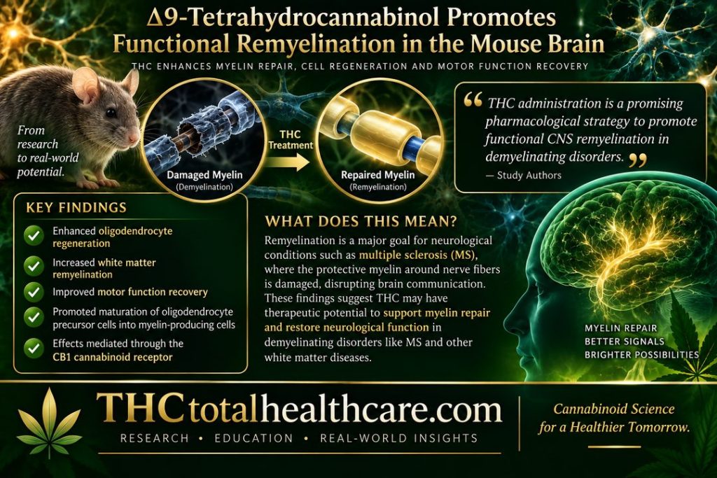
Δ 9 -Tetrahydrocannabinol promotes functional remyelination in the mouse brain

Myelin is the protective coating that surrounds nerve fibers and helps electrical signals move efficiently through the brain and spinal cord. Damage to myelin is a major feature of neurological disorders involving demyelination, making the repair of this protective layer an important therapeutic target.

In this mouse study, Δ9-THC promoted the regeneration of oligodendrocytes, the cells responsible for producing myelin, and enhanced remyelination in damaged white matter. THC treatment was also associated with improved recovery of motor function.

The researchers found that these effects involved activation of the CB1 cannabinoid receptor and the maturation of oligodendrocyte precursor cells into myelin-producing cells. The findings suggest that THC may have potential as a compound capable of supporting functional repair after myelin damage.

“Background and purpose: Research on demyelinating disorders aims to find novel molecules that are able to induce oligodendrocyte precursor cell differentiation to promote central nervous system remyelination and functional recovery. Δ9 -Tetrahydrocannabinol (THC), the most prominent active constituent of the hemp plant Cannabis sativa, confers neuroprotection in animal models of demyelination. However, the possible effect of THC on myelin repair has never been studied.

Experimental approach: By using oligodendroglia-specific reporter mouse lines in combination with two models of toxin-induced demyelination, we analysed the effect of THC on the processes of oligodendrocyte regeneration and functional remyelination.

Key results: We show that THC administration enhanced oligodendrocyte regeneration, white matter remyelination and motor function recovery. THC also promoted axonal remyelination in organotypic cerebellar cultures. THC remyelinating action relied on the induction of oligodendrocyte precursor differentiation upon cell cycle exit and via CB1 cannabinoid receptor activation.

Conclusions and implications: Overall, our study identifies THC administration as a promising pharmacological strategy aimed to promote functional CNS remyelination in demyelinating disorders.”

https://pubmed.ncbi.nlm.nih.gov/34216154/

“THC administration following cuprizone-induced demyelination enhanced oligodendrocyte regeneration, white matter remyelination and motor function recovery.”

“THC administration is a promising pharmacological strategy to promote functional CNS remyelination in demyelinating disorders.”

“Our study provides a novel therapeutic advantage of THC-based interventions in multiple sclerosis by promoting remyelination and functional recovery. New clinical trials with improved designs on cannabinoids in people with multiple sclerosis are needed now, considering these compounds as potential remyelinating/disease-modifying drugs to try to overcome previous failures. Our work also suggests that at least part of the neuroprotective action of phytocannabinoids in multiple sclerosis animal models and potentially in patients as well may be due to an enhanced CNS remyelination. Finally, this study also identifies THC as a potent inductor of oligodendrocyte progenitor cell differentiation under demyelination in mice, opening the possibility for this molecule to become a candidate drug to promote oligodendrocyte regeneration and remyelination in the treatment of demyelinating disorders.”

https://bpspubs.onlinelibrary.wiley.com/doi/10.1111/bph.15608

Need this THC study explained? thcXplained.com

CBG, CBD, Δ9-THC, CBN, CBGA, CBDA and Δ9-THCA as antioxidant agents and their intervention abilities in antioxidant action

Fitoterapia“Positive effect of some cannabinoids in the treatment and prophylaxis of a wide variety of oxidation-associated diseases and growing popularity of supplements containing cannabinoids, mainly cannabinoid oils (e.g. CBD oil, CBG oil), in the self-medication of humans cause a growing interest in the antioxidant properties of these compounds, especially those not showing psychotropic effects.

Herein, we report the antioxidant activity of cannabigerol (CBG), cannabidiol (CBD), Δ9-tetrahydrocannabinol (Δ9-THC), cannabinol (CBN), cannabigerolic acid (CBGA), cannabinolic acid (CBDA) and Δ9-tetrahydrocannabinolic acid (Δ9-THCA) estimated by spectrophotometric methods: ABTS, DPPH, ORAC, beta-carotene CUPRAC and FRAP.

The presented data prove that all the examined cannabinoids exhibit antioxidant activity manifested in their ability to scavenge free radicals, to prevent the oxidation process and to reduce metal ions. Although the intensity of these activities is not the same for the individual cannabinoids it is comparable for all of them with that of E vitamin.”

https://pubmed.ncbi.nlm.nih.gov/33964342/

“The present paper discusses the antioxidant properties of CBG, CBN, CBDA, CBGA and Δ9-THCA which, beside CBD and Δ9-THC, are also supposed to be bioactive compounds useful in the therapeutic treatment of different diseases. According to the literature, CBD and Δ9-THC exhibit strong antioxidant activity, stronger than vitamins C, A and E.

The presented data prove that all the examined cannabinoids – CBG, CBD, Δ9-THC, CBN, CBGA CBDA and Δ9-THCA – exhibit antioxidant activity manifesting itself in their ability to scavenge free radicals, to protect oxidation process and to reduce metal ions. Although, the intensity of these activities for individual cannabinoids is not the same, it is generally comparable to that of E vitamin.”  https://www.sciencedirect.com/science/article/pii/S0367326X21000903?via%3Dihub