Combined Cannabidiol and Δ9-Tetrahydrocannabinol Modulate Inflammation and Nociceptive Signaling in Human Dental Pulp Cells

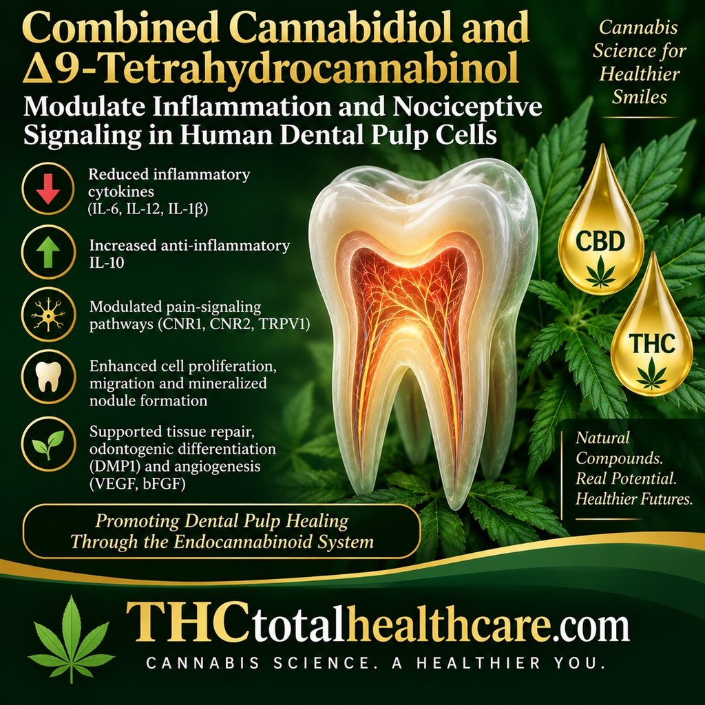
A new study using human dental pulp cells found that combining cannabidiol (CBD) and Δ9-tetrahydrocannabinol (THC) produced stronger anti-inflammatory effects than either cannabinoid alone. The combination reduced the pro-inflammatory cytokines IL-6, IL-12, and IL-1β while increasing the anti-inflammatory cytokine IL-10.

CBD and THC together also influenced important pathways involved in pain signaling and healing. Researchers reported increased expression of the cannabinoid receptors CNR1 and CNR2, modulation of TRPV1, and enhanced cell proliferation and migration. The combination also promoted mineralized nodule formation and increased markers associated with odontogenic differentiation and new blood-vessel formation.

The researchers concluded that combined CBD and THC show promise for promoting dental pulp healing by simultaneously influencing inflammation, nociceptive signaling, tissue repair, angiogenesis, and cell migration. The findings provide new evidence that cannabinoids may have potential applications in future vital pulp therapies designed to preserve and regenerate living dental tissue.

“Introduction: Dental pulp inflammation, caused by caries, trauma, or restorative operations, produces pain and slows regeneration. Cannabidiol (CBD) and Δ9-tetrahydrocannabinol (THC), cannabinoids of Cannabis sativa, have effects on inflammation and pain relief. However, the impact of these factors on dental pulp healing is not understood. This study investigated the combined effects of CBD and THC in controlling inflammation and nociception during dental pulp repair.

Methods: Human dental pulp cells were stimulated with lipopolysaccharide (LPS) and treated with CBD (2.5 μg/mL), THC (0.6 μg/mL), and their combination. Pro-inflammatory cytokines (IL-6, IL-12 and IL-1β) and anti-inflammatory cytokine IL-10 were analysed using qPCR and ELISA, while anti-nociceptive markers (CNR1, CNR2, and TRPV1) were evaluated by qPCR and Western blot. Cell viability, proliferation, and migration assays were performed to evaluate treatment effects on cellular behaviours associated with pulpal repair and regenerative potential.

Results: Compared with either treatment alone, the CBD/THC combination more effectively attenuated pro-inflammatory cytokines IL-6, IL-12, and IL-1β, enhanced CNR1 and CNR2 expression, modulated TRPV1, and increased IL-10 expression. Viability assays determined optimal non-cytotoxic concentrations for treatment. The combination enhanced cell proliferation and migration, promoted mineralized nodule formation, and significantly upregulated markers associated with tissue repair, odontogenic differentiation (DMP1), and angiogenesis (VEGF and bFGF).

Conclusion: Combined CBD and THC show promise for promoting dental pulp healing by modulating inflammation and nociceptive signaling, while supporting odontogenic differentiation, angiogenesis, and cell migration in human dental pulp cells. These findings provide in vitro evidence supporting combined cannabinoid treatment as a therapeutic strategy for vital pulp therapies.”

https://pubmed.ncbi.nlm.nih.gov/42722230

https://www.jendodon.com/article/S0099-2399(26)00500-5/abstract
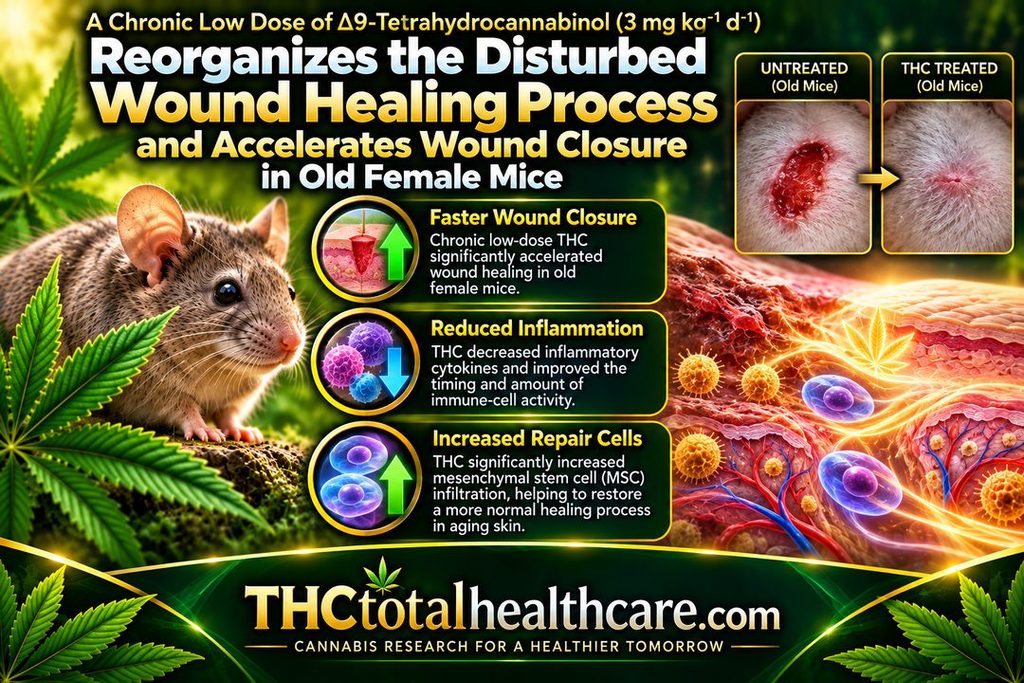
A chronic low dose of Δ9-tetrahydrocannabinol (3 mg / kg / 21 d) reorganizes the disturbed wound healing process and accelerates wound closure in old female mice

A chronic low dose of Δ9-THC accelerated wound closure in old female mice and helped reorganize an age-disrupted healing process. The biggest improvement occurred during the inflammatory phase, when aged wounds normally heal more slowly and remain inflamed longer.

THC changed the timing and amount of immune-cell activity in the wound, reduced inflammatory cytokines, and significantly increased infiltration of mesenchymal stem cells (MSCs) involved in tissue repair. The treatment did not increase growth-factor release, suggesting the benefit came mainly from better coordination of inflammation and cell migration.

The researchers concluded that THC improved wound healing by restoring a more normal sequence of immune-cell infiltration and cytokine release. The findings suggest that stimulating the aging endocannabinoid system may help support skin regeneration and tissue repair in older organisms.

“Wound healing in old mice is characterized by disturbed tissue homeostasis, manifested by delayed immune cell infiltration and reduced growth factor secretion, leading to a delayed onset and prolonged duration of the inflammatory phase.

The endocannabinoid system (ECS) is an important regulator of tissue homeostasis and cell migration and is also considered to be subject to aging processes, which may contribute to observable aging phenomena. Therefore, stimulating the aged ECS could represent a therapeutic option to support tissue regeneration in aging.

Female old mice received a low-dose of medical THC daily for 3 weeks, before four excisional full skin wounds were created. At day 1, 3 and 7 post-surgery, the wound closure rate was analyzed and wound samples were examined immunohistochemically for the numbers of granulocytes, M1-macrophages and mesenchymal stem cells (MSCs). The concentrations of inflammatory cytokines and regenerative growth factors were determined by ELISA.

Administration of THC improved the wound healing rate of old mice between day 1 and 7, which was associated with an altered timing and quantity of infiltrating immune cells and decreased levels of inflammatory cytokines in wound tissue on days 1 and 3 post-injury.

THC treatment significantly increased MSC infiltration but had no effect on the growth factor release.

The present study confirmed the anti-inflammatory activity of THC in vivo.

The THC-treatment improved wound healing in old mice by coordinating the temporal sequence of immune cell infiltration and cytokine release. Thus, restoration of ECS signaling could be an effective strategy to support age-related skin regeneration.”

https://pubmed.ncbi.nlm.nih.gov/40653209/

“A chronic low dose of THC accelerates the wound closure rate during the inflammatory phase in old mice.”

https://www.sciencedirect.com/science/article/pii/S0531556525001615?via%3Dihub

Meed this THC study explained? thcXplained.com

∆9-Tetrahydrocannabinol Increases Growth Factor Release by Cultured Adipose Stem Cells and Adipose Tissue in vivo

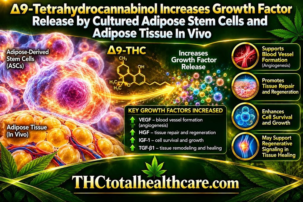
Δ9-THC increased the release of growth factors from cultured adipose-derived stem cells and from adipose tissue in vivo. These growth factors are involved in processes such as cell survival, tissue repair, blood-vessel formation, and regeneration.

The researchers found that THC changed the secretory activity of adipose stem cells rather than simply affecting their growth. This suggests that THC may influence how these cells communicate with surrounding tissue and support repair through the factors they release.

The findings point to a potential role for THC in regenerative signaling and tissue-healing biology, particularly through adipose-derived stem cells and the local tissue environment. The study adds another dimension to cannabinoid research by showing that THC can affect not just mature cells, but also the reparative signals produced by stem-cell populations.

“Background: Because of its biocompatibility and its soft and dynamic nature, the grafting of adipose tissue is regarded an ideal technique for soft-tissue repair. The adipose stem cells (ASCs) contribute significantly to the regenerative potential of adipose tissue, because they can differentiate into adipocytes and release growth factors for tissue repair and neovascularization to facilitate tissue survival. The present study tested the effect of administering a chronic low dose of ∆9-tetrahydrocannabinol (THC) on these regenerative properties, in vitro and in vivo.

Methods: Human ASCs were exposed to increasing concentrations of THC. Resazurin conversion was applied to investigate the effect on metabolic activity, cell number was assessed by crystal violet staining, tri-linear differentiation was evaluated by specific colorimetric approaches, and the release of growth factors was analyzed by ELISA. Two groups of mice were treated daily either with a low dose of THC (3 mg/kg) or a vehicle solution. After 3 weeks, adipose tissue was obtained from excised fat deposits, homogenized and tested for growth factor contents.

Results: THC decreased ASC proliferation but increased metabolic activity as well as adipogenic and chondrogenic differentiation. A low concentration of THC (1 µM) enhanced the growth factor release by ASCs. The concentration of these cytokines was also increased in adipose tissue of mice treated with THC.

Conlusion: Our results indicate that chronic activation of the endocannabinoid system promoted differentiation and growth factor release of ASCs, which could be of specific value for enhancing the regenerative potential of adipose tissue.”

https://pubmed.ncbi.nlm.nih.gov/39825991/

“Our findings may offer a new approach for improved application of adipose tissue in regenerative medicine.”

https://link.springer.com/article/10.1007/s13770-024-00692-8

Need this THC study explained? thcXplained.com