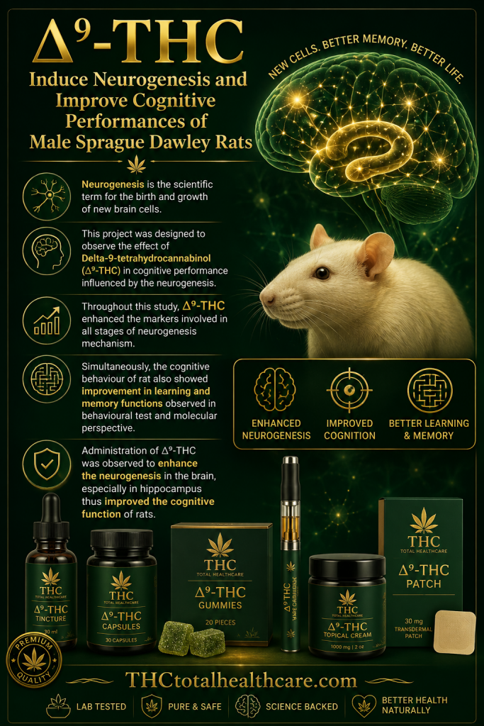
Delta-9-Tetrahydrocannabinol (∆9-THC) Induce Neurogenesis and Improve Cognitive Performances of Male Sprague Dawley Rats

THC is often associated with impaired memory, but its effects on the brain may depend heavily on dose and context. In this rat study, Δ9-THC enhanced markers involved in multiple stages of hippocampal neurogenesis and improved learning and memory performance, with the 1.5 mg/kg dose producing particularly strong effects on both neurogenesis and cognitive function.

“Neurogenesis is influenced by various external factors such as enriched environments. Some researchers had postulated that neurogenesis has contributed to the hippocampal learning and memory.

This project was designed to observe the effect of Delta-9-tetrahydrocannabinol (∆9-THC) in cognitive performance that influenced by the neurogenesis.

Different doses of ∆9-THC were used for observing the neurogenesis mechanism occurs in the hippocampus of rats. The brains were stained with antibodies, namely BrdU, glial fibrillary acidic protein (GFAP), nestin, doublecortin (DCX) and class III β-tubulin (TuJ-1). The cognitive test was used novel-object discrimination test (NOD) while the proteins involved, DCX and brain-derived neurotrophic factor (BDNF), were measured.

Throughout this study, ∆9-THC enhanced the markers involved in all stages of neurogenesis mechanism. Simultaneously, the cognitive behaviour of rat also showed improvement in learning and memory functions observed in behavioural test and molecular perspective.

Administration of ∆9-THC was observed to enhance the neurogenesis in the brain, especially in hippocampus thus improved the cognitive function of rats.”

https://pubmed.ncbi.nlm.nih.gov/28933048

“The treatment of 1.5 mg/kg of ∆9-THC has increase all the markers for neurogenesis and cognition function while improve the cognitive performance.”

https://link.springer.com/article/10.1007/s12640-017-9806-x

“Neurogenesis is the scientific term for the birth and growth of new brain cells.”

Need this THC study explained? thcXplained.com
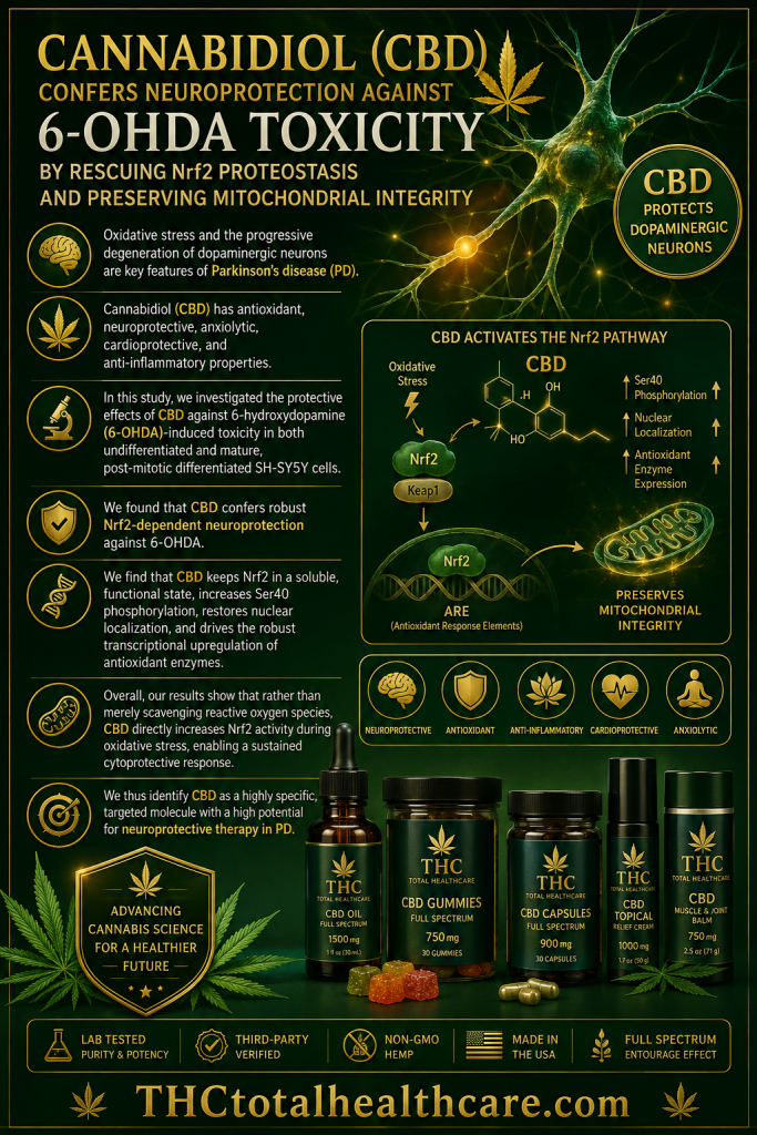
Cannabidiol confers neuroprotection against 6-OHDA toxicity by rescuing Nrf2 proteostasis and preserving mitochondrial integrity

Oxidative stress and mitochondrial dysfunction are major contributors to the loss of dopamine-producing neurons in Parkinson’s disease. In this cellular model, cannabidiol (CBD) protected against 6-OHDA-induced neurotoxicity by restoring Nrf2-dependent antioxidant defenses, preserving mitochondrial integrity, and reducing cellular damage.

“Oxidative stress and the progressive degeneration of dopaminergic neurons are key features of Parkinson’s disease (PD). The intrinsically disordered structure of the transcription factor Nuclear factor erythroid 2-related factor 2 (Nrf2), which coordinates the main cellular antioxidant response of the body, makes it highly susceptible to misfolding and aggregation under severe oxidative stress, compromising cellular survival.

Cannabidiol (CBD) has potent neuroprotective properties, but its exact molecular mechanism within the dopaminergic redox environment remains unclear. In this study, we investigated the protective effects of CBD against 6-hydroxydopamine (6-OHDA)-induced toxicity in both undifferentiated and mature, post-mitotic differentiated SH-SY5Y cells.

We found that CBD confers robust Nrf2-dependent neuroprotection against 6-OHDA. Importantly, we uncover a previously unexplored mechanism of neuroprotection by which CBD actively prevents the stress-induced sequestration of Nrf2 into insoluble cytoplasmic inclusions under oxidative stress.

We find that CBD keeps Nrf2 in a soluble, functional state, increases Ser40 phosphorylation, restores nuclear localization, and drives the robust transcriptional upregulation of antioxidant enzymes. This targeted activation of Nrf2 effectively reduces intracellular reactive oxygen species (ROS), significantly attenuates mitochondrial fragmentation, and decreases aberrant mitophagic activity.

Overall, our results show that rather than merely scavenging reactive oxygen species, CBD directly increases Nrf2 activity during oxidative stress, enabling a sustained cytoprotective response.

We thus identify CBD as a highly specific, targeted molecule with a high potential for neuroprotective therapy in PD.”

https://pubmed.ncbi.nlm.nih.gov/42570822

“CBD has antioxidant, neuroprotective, anxiolytic, cardioprotective, and anti-inflammatory properties.”

https://www.sciencedirect.com/science/article/pii/S0891584926010105?via%3Dihub


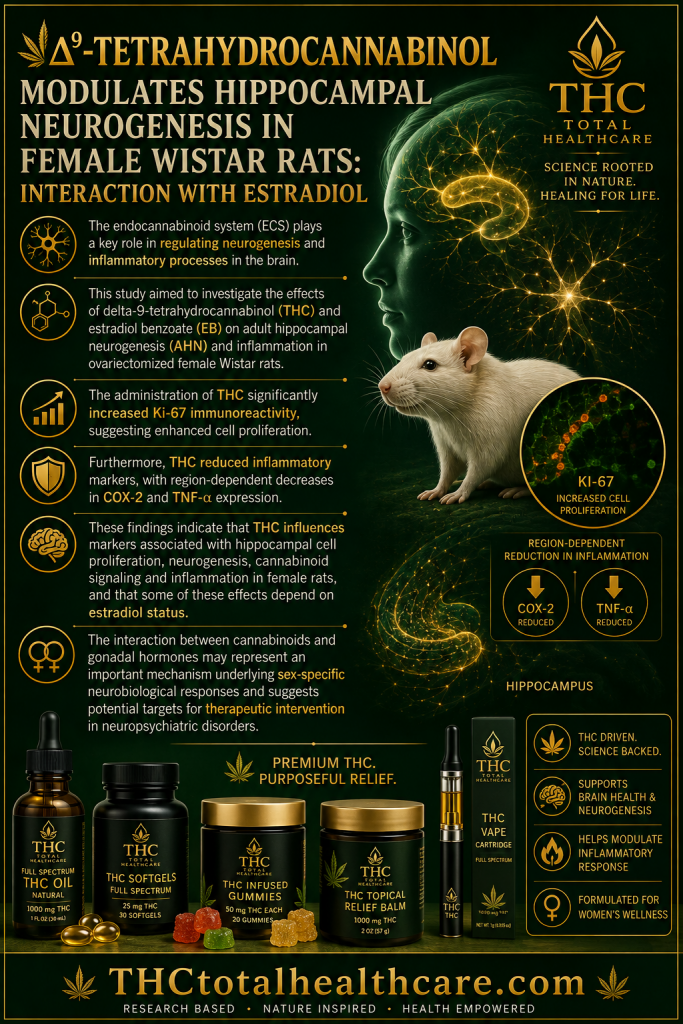
Δ9-Tetrahydrocannabinol Modulates Hippocampal Neurogenesis in Female Wistar Rats: Interaction with Estradiol

THC may influence the female brain through both neurogenic and anti-inflammatory mechanisms. In ovariectomized female rats, Δ9-THC significantly increased hippocampal cell proliferation, showed evidence of enhanced neurogenesis, and reduced the inflammatory markers COX-2 and TNF-α. The effects also interacted with estradiol, highlighting the importance of hormonal status in determining THC’s actions in the brain.

“The endocannabinoid system (ECS) plays a key role in regulating neurogenesis and inflammatory processes in the brain.

The increasing prevalence of Cannabis use among women highlights the importance of understanding sex-specific effects of cannabinoids, particularly in the context of hormonal interactions.

This study aimed to investigate the effects of delta-9-tetrahydrocannabinol (THC) and estradiol benzoate (EB) on adult hippocampal neurogenesis (AHN) and inflammation in ovariectomized female Wistar rats.

Sixteen rats were allocated to four experimental groups receiving THC, EB, both treatments, and vehicle. Immunohistochemical analyses were conducted to evaluate markers of proliferation (Ki-67), neurogenesis (doublecortin and PSA-NCAM), cannabinoid receptor expression (CB1), and inflammation (COX-2 and TNF-α) in the hippocampal formation.

The administration of THC significantly increased Ki-67 immunoreactivity, suggesting enhanced cell proliferation. A trend toward increased doublecortin expression was observed, particularly in EB-treated animals. THC also modulated CB1 receptor expression, with significant increases in the dentate gyrus and hilus following combined THC and EB treatment. Furthermore, THC reduced inflammatory markers, with region-dependent decreases in COX-2 and TNF-α expression.

These findings indicate that THC influences markers associated with hippocampal cell proliferation, neurogenesis, cannabinoid signaling and inflammation in female rats, and that some of these effects depend on estradiol status.

The interaction between cannabinoids and gonadal hormones may represent an important mechanism underlying sex-specific neurobiological responses and suggests potential targets for therapeutic intervention in neuropsychiatric disorders.”

https://pubmed.ncbi.nlm.nih.gov/42570151

https://link.springer.com/article/10.1007/s11064-026-04857-w


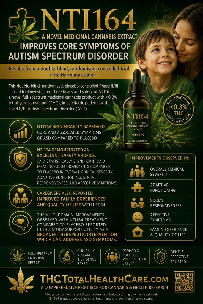
NTI164, a novel medicinal cannabis extract, improves core symptoms of autism spectrum disorder: Results from a double-blind, randomised, controlled trial (The Harmony study)

Current medications for autism spectrum disorder generally target associated symptoms rather than the core features of ASD. In this randomized, double-blind, placebo-controlled Phase II/III trial, the full-spectrum medicinal cannabis extract NTI164 significantly improved overall clinical severity, adaptive functioning, social responsiveness, mood, anxiety, and family quality of life in children and adolescents with moderate-to-severe ASD, while demonstrating an excellent safety profile.

“Autism spectrum disorder (ASD) is a heterogeneous neurodevelopmental disorder, characterised by difficulties with communication, social interaction, repetitive behaviours, restricted interests, and varying levels of intellectual disability. Aetiology remains unclear for many patients and the underlying physiology is complex. Approved pharmacological treatments of ASD target irritability, temper tantrums, and agitation, with no therapies targeting core ASD symptoms.

This double-blind, randomised, placebo-controlled Phase II/III clinical trial investigated the efficacy and safety of NTI164, a novel full-spectrum medicinal cannabis product with <0.3% tetrahydrocannabinol (THC), in paediatric patients with Level II/III ASD.

Participants were recruited from a tertiary paediatric neurology clinic and randomised to receive NTI164 up to 20 mg/kg/day or placebo for an 8-week double-blind phase; participants receiving placebo were able to receive NTI164 in an 8-week open label phase following the double-blind phase. Safety assessments, clinician-, and caregiver-rated tools measuring symptoms were utilised at baseline and Week 8. Analysis of Covariance (ANCOVA) was used for statistical analyses.

NTI164 demonstrated an excellent safety profile, and statistically significant and meaningful improvements compared to placebo in overall clinical severity, adaptive functioning, social responsiveness, and affective symptoms. Caregivers also reported improved family experiences and quality of life with NTI164. Participants who transitioned from placebo to NTI164 open label reported similar improvements as those reported during the double-blind phase.

NTI164 significantly improved core and associated symptoms of ASD compared to placebo.

Consistent benefits reported by both clinicians and caregivers in both open label and double-blind contexts supports further clinical development of NTI164 in ASD.”

https://pubmed.ncbi.nlm.nih.gov/42561512

“The multi-domain improvements observed with NTI164 treatment compared to placebo reported in this study support utility as a broader therapeutic intervention which can address ASD symptoms.

This randomised, double-blind, placebo-controlled clinical trial in children and adolescents with moderate-severe ASD showed NTI164 meaningfully improves global impression of clinical severity, adaptive functioning, aspects of social responsiveness, mood, anxiety, and family experiences, with an excellent safety profile.”

https://www.neurotherapeuticsjournal.org/article/S1878-7479(26)00203-5/fulltext


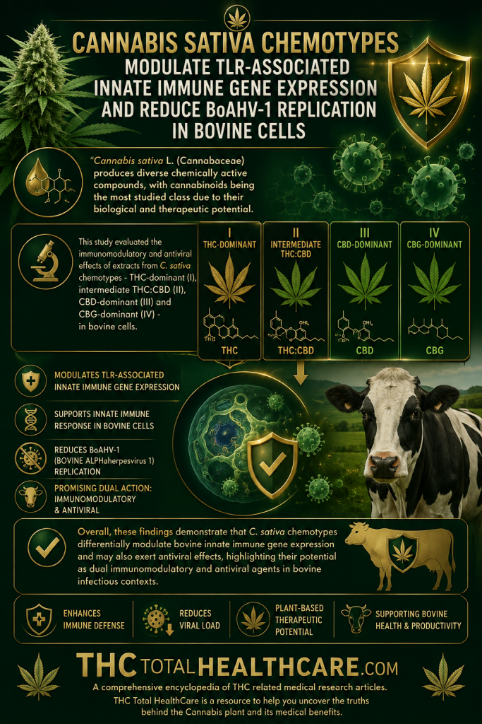
Cannabis sativa chemotypes modulate TLR-associated innate immune gene expression and reduce BoAHV-1 replication in bovine cells

Cannabis compounds may influence antiviral defenses as well as inflammation. In bovine cells infected with bovine alphaherpesvirus 1, selected Cannabis sativa chemotypes altered TLR-associated innate immune gene expression and reduced viral replication, suggesting that cannabis-derived compounds may have broader immunomodulatory and antiviral potential worth further investigation.

“Cannabis sativa L. produces a wide range of bioactive metabolites, including phytocannabinoids such as tetrahydrocannabinol (THC), cannabidiol (CBD) and cannabigerol (CBG), which exhibit antiviral activity and modulate innate immune responses through Toll-like receptors (TLRs), particularly TLR4 and TLR7. The cross-regulation between cannabinoids and TLRs can influence the production of cytokines and antimicrobial peptides.

Given their key role in orchestrating innate immunity, particularly inflammatory responses and antiviral activity, understanding these processes in bovine immune cells is essential.

This study evaluated the immunomodulatory and antiviral effects of extracts from C. sativa chemotypes – THC-dominant (I), intermediate THC:CBD (II), CBD-dominant (III) and CBG-dominant (IV) – in bovine cells.

In peripheral blood mononuclear cells, chemotype I induced an enhanced inflammatory response, increasing TLR4 and BMAP28 transcription and pro-inflammatory cytokine expression at both transcriptional and protein levels. Similarly, chemotype IV promoted a pro-inflammatory profile characterised by increased TLR4, BMAP28 and IFNβ expression, as well as elevated TNFα protein levels.

In contrast, chemotypes II and III elicited anti-inflammatory effects. Chemotype III decreased TLR4, TLR7, BMAP28, TNFα and IFNβ transcription, although IFNγ protein levels increased. Chemotype II produced a comparable, although less pronounced, anti-inflammatory pattern, reducing TLR4, TLR7, TNFα and IFNβ while increasing BMAP28. Additionally, chemotypes II, III, and IV exhibited antiviral activity in BoAHV-1-infected MDBK cells, significantly reducing viral titres at 48 h post-infection.

Overall, these findings demonstrate that C. sativa chemotypes differentially modulate bovine innate immune gene expression and may also exert antiviral effects, highlighting their potential as dual immunomodulatory and antiviral agents in bovine infectious contexts.”

https://pubmed.ncbi.nlm.nih.gov/42561569

Cannabis sativa L. (Cannabaceae) produces diverse chemically active compounds, with cannabinoids being the most studied class due to their biological and therapeutic potential.”

https://www.sciencedirect.com/science/article/abs/pii/S0161589026001768?via%3Dihub


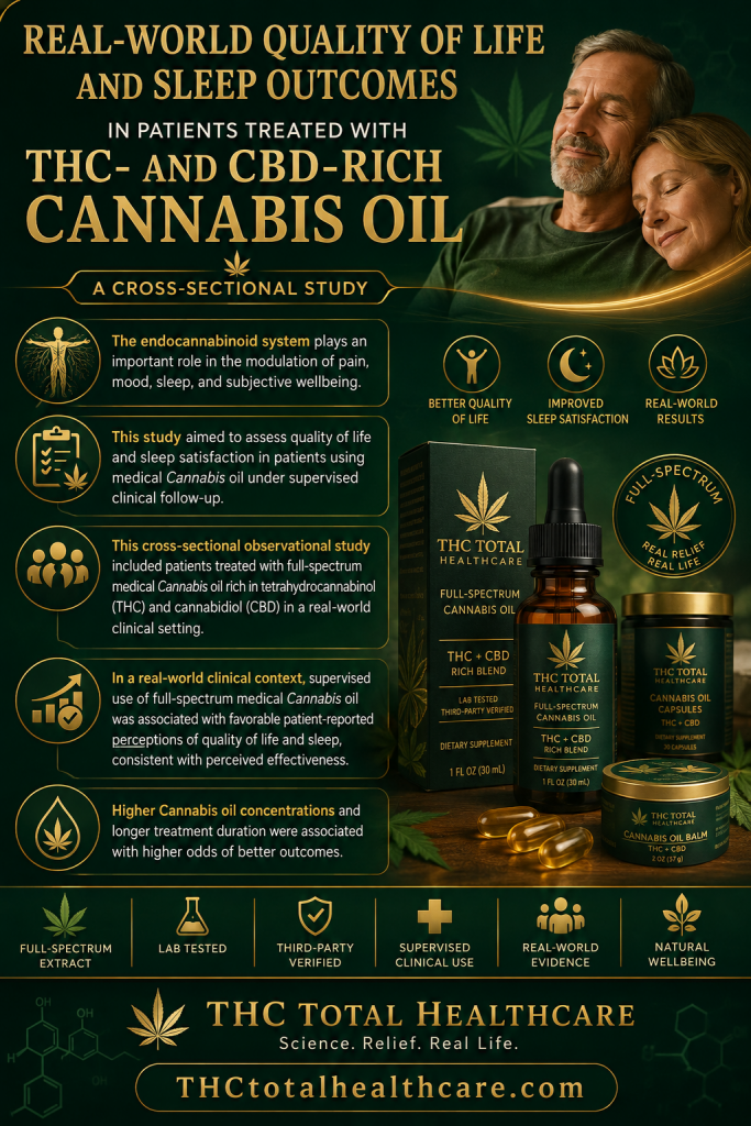
Real-world quality of life and sleep outcomes in patients treated with THC- and CBD-rich Cannabis oil: a cross-sectional study

Patients using THC- and CBD-rich cannabis oil reported meaningful improvements in both sleep and overall quality of life in this real-world study. The findings suggest that cannabinoid-rich oil formulations may help address multiple symptom domains at once, particularly in patients dealing with persistent sleep disturbance and reduced day-to-day well-being.

“The endocannabinoid system plays an important role in the modulation of pain, mood, sleep, and subjective wellbeing. Despite the growing clinical use of medicinal Cannabis, real-world data simultaneously evaluating quality of life and sleep-related outcomes in heterogeneous clinical populations remain limited.

This study aimed to assess quality of life and sleep satisfaction in patients using medical Cannabis oil under supervised clinical follow-up. This cross-sectional observational study included patients treated with full-spectrum medical Cannabis oil rich in tetrahydrocannabinol (THC) and cannabidiol (CBD) in a real-world clinical setting.

Quality of life was assessed using the World Health Organization Quality of Life-BREF (WHOQOL-BREF), and sleep quality was evaluated using the Pittsburgh Sleep Quality Index (PSQI). Sociodemographic, clinical, and treatment-related data were collected via an electronic questionnaire. Nonparametric analyses, Spearman correlation, and ordinal logistic regression models were performed. Seventy-one participants were included, predominantly female, with diverse clinical conditions.

Participants reported generally favorable perceptions regarding quality of life and sleep satisfaction, with median scores concentrated in the higher response categories. Positive correlations were observed between the psychological domains of quality of life and sleep satisfaction.

Higher Cannabis oil concentrations and longer treatment duration were associated with higher odds of better outcomes.

In a real-world clinical context, supervised use of full-spectrum medical Cannabis oil was associated with favorable patient-reported perceptions of quality of life and sleep, consistent with perceived effectiveness.

These findings highlight the need for longitudinal studies with pre-treatment baseline assessment.”

https://pubmed.ncbi.nlm.nih.gov/42558508

Cannabis sativa L. [Cannabaceae] has a documented history of medicinal use spanning thousands of years across diverse cultural and geographic contexts, with traditional applications encompassing pain relief, mood modulation, sleep induction, and the management of inflammatory and neurological conditions.”

“The growing integration of Cannabis into contemporary clinical practice reflects both the long-standing ethnopharmacological tradition associated with this species and the expanding body of evidence supporting the pharmacological activity of its principal phytocannabinoid metabolites, tetrahydrocannabinol (THC) and cannabidiol (CBD).”

“In summary, the results suggest that, in supervised clinical practice, patients using full-spectrum THC and CBD rich oil reported favorable perceived quality of life and sleep satisfaction.”

https://www.frontiersin.org/journals/pharmacology/articles/10.3389/fphar.2026.1862725/full


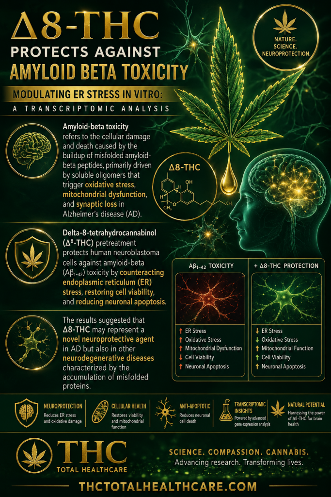
Δ8-THC Protects against Amyloid Beta Toxicity Modulating ER Stress In Vitro: A Transcriptomic Analysis

Amyloid-beta toxicity is a major driver of neuronal stress and degeneration in Alzheimer’s disease. In this laboratory study, Δ8-THC protected cells against amyloid-beta-induced damage and altered gene-expression pathways involved in endoplasmic reticulum stress. The results demonstrated that Δ8-THC might represent a new neuroprotective agent in Alzheimer’s disease.

“Alzheimer’s disease (AD) represents the most common form of dementia, characterized by amyloid β (Aβ) plaques and neurofibrillary tangles (NFTs). It is characterized by neuroinflammation, the accumulation of misfolded protein, ER stress and neuronal apoptosis. It is of main importance to find new therapeutic strategies because AD prevalence is increasing worldwide.

Cannabinoids are arising as promising neuroprotective phytocompounds.

In this study, we evaluated the neuroprotective potential of Δ8-THC pretreatment in an in vitro model of AD through transcriptomic analysis.

We found that Δ8-THC pretreatment restored the loss of cell viability in retinoic acid-differentiated neuroblastoma SH-SY5Y cells treated with Aβ1-42. Moreover, the transcriptomic analysis provided evidence that the enriched biological processes of gene ontology were related to ER functions and proteostasis. In particular, Aβ1-42 upregulated genes involved in ER stress and unfolded protein response, leading to apoptosis as demonstrated by the increase in Bax and the decrease in Bcl-2 both at gene and protein expression levels. Moreover, genes involved in protein folding and degradation were also deregulated. On the contrary, Δ8-THC pretreatment reduced ER stress and, as a consequence, neuronal apoptosis.

Then, the results demonstrated that Δ8-THC might represent a new neuroprotective agent in AD.”

https://pubmed.ncbi.nlm.nih.gov/37047608

“The results suggested that Δ8-THC may represent a novel neuroprotective agent in AD but also in other neurodegenerative diseases characterized by the accumulation of misfolded proteins.”

https://www.mdpi.com/1422-0067/24/7/6598

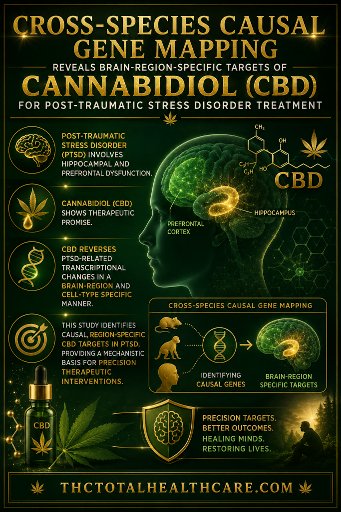
Cross-species causal gene mapping reveals brain-region-specific targets of Cannabidiol for post-traumatic stress disorder treatment

Post-traumatic stress disorder involves complex changes across multiple brain regions, and cannabidiol (CBD) has emerged as a potential treatment candidate. Using cross-species causal gene mapping, researchers found that CBD reverses PTSD-related transcriptional changes in a brain-region and cell-type specific manner, while also identifying molecular targets that may help explain its therapeutic effects and guide future PTSD treatment development.

Purpose: Post-traumatic stress disorder (PTSD) involves hippocampal and prefrontal dysfunction. Cannabidiol (CBD) shows therapeutic promise, but its cell-type-specific and causal mechanisms remain unclear.

Methods: We performed single-cell RNA sequencing of hippocampal and prefrontal cortices from PTSD mice, healthy controls, and CBD-treated PTSD mice. Genes dysregulated in PTSD and reversed by CBD were integrated with human PTSD GWAS and brain eQTL datasets using summary-data-based Mendelian randomization (SMR) to identify causal risk targets. Molecular docking assessed direct CBD-protein interactions.

Results: PTSD induced extensive transcriptional alterations, most prominent in excitatory neurons. SMR analysis identified 15 potential causal risk genes linked to PTSD. Among these, 7 high-confidence targets were confirmed to be transcriptionally responsive to CBD treatment. In the hippocampus, key causal targets included LYNX1 (OR = 0.75, 95% CI: 0.57-0.98, P = 0.038), RAB3C (OR = 1.291, 95% CI: 1.052-1.584, P = 0.015), MAGI2 (OR = 1.15, 95% CI: 1.00-1.31, P = 0.048), LINGO2 (OR = 1.48, 95% CI: 1.07-2.03, P = 0.017), and UNC5D (OR = 1.41, 95% CI: 1.03-1.93, P = 0.032). In the prefrontal cortex, identified targets were CNTN3 (OR = 0.78, 95% CI: 0.62-0.98, P = 0.036), IGSF21 (OR = 1.22, 95% CI: 1.04-1.42, P = 0.012). While SEPTIN3 (OR = 1.25, 95% CI: 1.01-1.56, P = 0.043) was identified as a causal risk gene, its expression was not reversed by CBD. Molecular docking indicated that all 15 SMR-identified candidates possess strong binding affinity to CBD, including RAB3C (-8.636 kcal/mol), CNTN3 (-7.216 kcal/mol), and LINGO2 (-6.222 kcal/mol), which suggests a direct pharmacological interaction.

Conclusion: This study identifies causal, region-specific CBD targets in PTSD, providing a mechanistic basis for precision therapeutic interventions.”

https://pubmed.ncbi.nlm.nih.gov/42542041

“CBD reverses PTSD-related transcriptional changes in a brain-region and cell-type specific manner.”

https://www.sciencedirect.com/science/article/abs/pii/S0006291X26011113?via%3Dihub

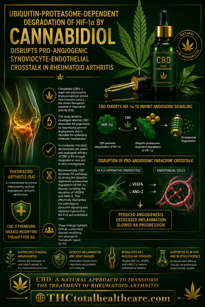
Ubiquitin-proteasome-dependent degradation of HIF-1α by cannabidiol disrupts pro-angiogenic synoviocyte-endothelial crosstalk in rheumatoid arthritis

Rheumatoid arthritis is driven in part by abnormal synovial inflammation and the formation of new blood vessels that sustain diseased tissue. In this study, cannabidiol (CBD) reduced joint inflammation, synovial angiogenesis, and bone destruction while suppressing abnormal fibroblast activity. The researchers concluded that CBD disrupts pro-angiogenic signaling through HIF-1α degradation and may represent a promising disease-modifying anti-angiogenic therapeutic agent for rheumatoid arthritis.

Background: Cannabidiol (CBD), a major non-psychoactive phytocannabinoid derived from Cannabis sativa L., has shown therapeutic potential in rheumatoid arthritis (RA). However, the mechanisms by which CBD modulates synovial angiogenesis remain unclear.

Purpose: This study aimed to investigate whether CBD attenuates RA progression by suppressing synovial angiogenesis and to elucidate the underlying molecular mechanisms.

Methods: An adjuvant-induced arthritis (AIA) rat model was established to evaluate the therapeutic effects of CBD in vivo using arthritis scoring, micro-CT, and histopathological, immunohistochemical, and immunofluorescence analyses. In vitro, cytotoxicity was determined using the CCK-8 assay, followed by evaluations of CBD’s direct effects on the proliferation, migration, invasion, and inflammatory responses of RA fibroblast-like synoviocytes (RA-FLS) were evaluated, alongside Cell Counting Kit-8 (CCK-8) for cytotoxicity screening. Furthermore, the paracrine regulation of angiogenesis was assessed using a conditioned medium (CM) transfer system from hypoxia-stimulated RA-FLS applied to human umbilical vein endothelial cells (HUVECs). Molecular mechanisms were analyzed via Western blotting, RT-qPCR, ELISA, co-immunoprecipitation (Co-IP), molecular docking, and targeted proteasome inhibition (MG132).

Results: In vivo, CBD (5 and 10 mg/kg) treatment markedly alleviated joint inflammation, synovial angiogenesis, and structural bone destruction in AIA rats. In vitro, non-cytotoxic concentrations of CBD (2.4-4.8 μM) significantly suppressed aberrant RA-FLS proliferation, migration, invasion, and pro-inflammatory cytokine secretion. Mechanistically, CBD abrogated the hypoxia-induced accumulation of hypoxia-inducible factor-1α (HIF-1α) protein in RA-FLS without significantly altering HIF1A mRNA expression. This reduction was effectively reversed by MG132. Co-IP and molecular docking analyses revealed that CBD directly enhances the polyubiquitination of HIF-1α through stable structural interactions, driving a ubiquitin-proteasome-dependent degradation mechanism. Consequently, CBD dose-dependently decreased the extracellular secretion of vascular endothelial growth factor A (VEGFA) and angiopoietin-2 (ANG-2) from RA-FLS. Functionally, CM from CBD-treated RA-FLS disrupted the pro-angiogenic paracrine crosstalk-independent of direct CBD carryover-significantly impairing HUVEC migration, capillary-like tube formation, and downstream VEGFR2 (Tyr1175) phosphorylation.

Conclusion: CBD attenuates RA pathogenesis not only by directly suppressing RA-FLS hyperactivity but also by severing the pro-angiogenic paracrine crosstalk between RA-FLS and endothelial cells. These effects are driven by the ubiquitin-proteasome-dependent degradation of HIF-1α in RA-FLS via direct structural engagement, which depletes VEGFA/ANG-2 production and subsequent endothelial VEGFR2 activation. These findings highlight CBD as a promising disease-modifying anti-angiogenic therapeutic agent for RA.”

https://pubmed.ncbi.nlm.nih.gov/42542056

“In conclusion, this study demonstrates the potent anti-angiogenic efficacy of CBD in RA through integrated in vivo and in vitro investigations. Mechanistically, CBD alleviates RA pathology by driving the ubiquitin-dependent proteasomal degradation of HIF-1α, thereby curtailing the secretion of VEGFA and ANG-2. This effectively dismantles the pathological paracrine signaling axis between hyperactive RA-FLS and endothelial cells.”

https://www.sciencedirect.com/science/article/abs/pii/S0944711326008925?via%3Dihub

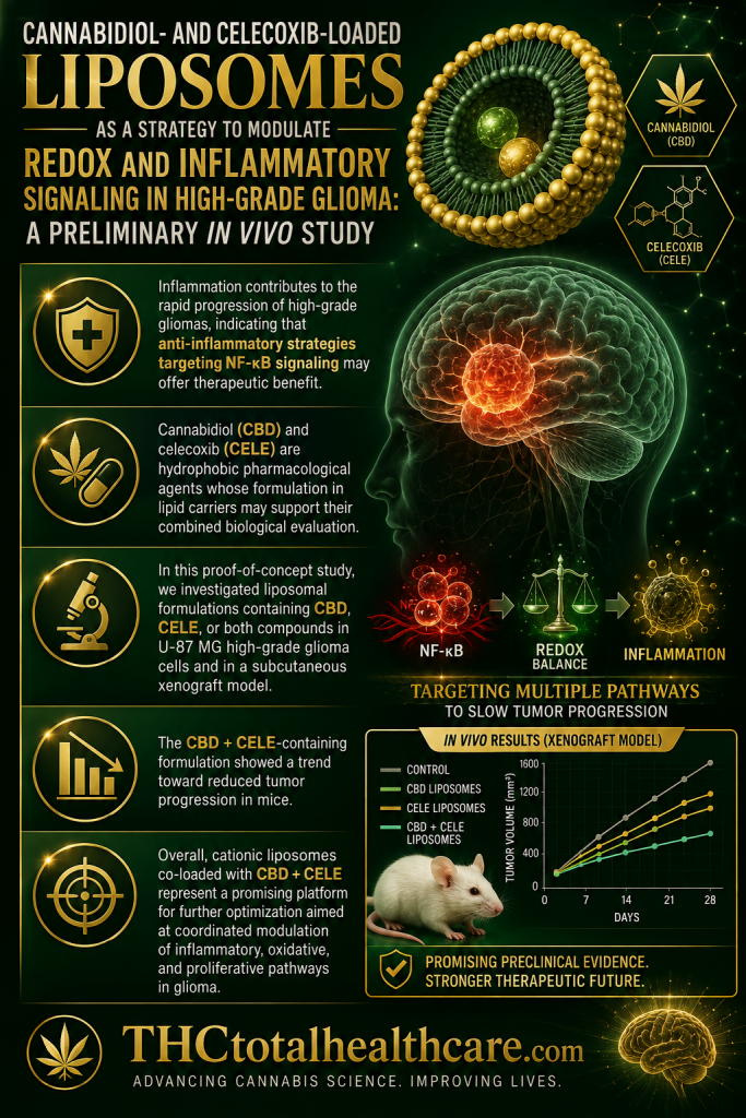
Cannabidiol- and Celecoxib-Loaded Liposomes as a Strategy to Modulate Redox and Inflammatory Signaling in High-Grade Glioma: A Preliminary In Vivo Study

High-grade glioma is marked by aggressive growth, inflammation, and oxidative stress, creating a need for therapies that can target several tumor-supporting pathways at once. In this preliminary in vivo study, liposomes carrying cannabidiol (CBD) and celecoxib modulated inflammatory and redox signaling in glioma tissue and showed antitumor effects. The researchers concluded that this combined liposomal approach may represent a promising strategy for further development against high-grade glioma.

“Inflammation contributes to the rapid progression of high-grade gliomas, indicating that anti-inflammatory strategies targeting NF-κB signaling may offer therapeutic benefit.

Cannabidiol (CBD) and celecoxib (CELE) are hydrophobic pharmacological agents whose formulation in lipid carriers may support their combined biological evaluation.

In this proof-of-concept study, we investigated liposomal formulations containing CBD, CELE, or both compounds in U-87 MG high-grade glioma cells and in a subcutaneous xenograft model.

We assessed cytotoxicity, apoptosis, oxidative stress, Nrf2-dependent responses, NF-κB-centered inflammatory networks, tumor cell invasive properties, and Wnt/β-catenin pathway activity. The nanoformulations induced reactive oxygen species generation by 1.8-fold, which was accompanied by Nrf2 activation. Cationic formulations loaded with the compounds produced more pronounced pro-apoptotic effects (up to 39%) than POPC liposomes, although both types reduced the nuclear translocation of the NF-κB p65 subunit.

The CBD + CELE-containing formulation showed a trend toward reduced tumor progression in mice. It is important to note that the in vitro and in vivo nanoformulations were physicochemically related, but not identical, and the in vivo experiment should be interpreted as a preliminary assessment after intratumoral administration.

Overall, cationic liposomes co-loaded with CBD + CELE represent a promising platform for further optimization aimed at coordinated modulation of inflammatory, oxidative, and proliferative pathways in glioma. However, additional studies, including tissue distribution, release kinetics, and efficacy in orthotopic glioma models, are needed to fully verify their translational potential.”

https://pubmed.ncbi.nlm.nih.gov/42511566

“In summary, this study shows that liposomal formulations containing CBD and CELE, particularly DOTAP:POPC formulations prepared with both compounds, modulate apoptosis, cell cycle distribution, oxidative stress, Nrf2/NF-κB/Wnt-related pathways, and glioma-associated inflammatory mediators in U-87 MG-based models. In the subcutaneous xenograft model, intratumoral administration of the CBD + CELE-containing formulation was associated with a trend toward reduced tumor progression and modulation of selected tumor-associated proteins.”

https://www.mdpi.com/1422-0067/27/14/6220