“Introduction: Metabolic dysfunction-associated steatotic liver disease (MASLD) is closely linked to alterations in liver lipid metabolism, oxidative stress, fibrosis, and dysregulation of the endocannabinoid system (ECS). Although increasing evidence supports a role for cannabinoids in metabolic disorders, most preclinical studies have been conducted in male models, leaving female-specific responses largely unexplored.
Methods: This study evaluated the effects of oral administration of a full-spectrum cannabis oil (CBD:THC 2:1) on MASLD-related alterations and ECS regulation in female Wistar rats fed a sucrose-rich diet (SRD). Rats were assigned to reference diet (RD), SRD, or SRD plus cannabis oil (1 mg/kg/day) for 3 weeks.
Results: SRD-fed rats developed liver steatosis and increased NAFLD activity score (NAS), accompanied by enhanced de novo lipogenesis, reduced mitochondrial fatty acid oxidation, increased oxidative stress, early fibrotic changes, and ECS overactivation. Cannabis oil administration improved liver lipid metabolism, reduced NAS and fibrosis markers, attenuated lipid peroxidation and oxidative stress, increased NrF2 and decreased NF-κB p65 expression, and normalized hepatic CB1 expression and circulating endocannabinoid levels.
Discussion: These findings demonstrate that full-spectrum cannabis oil is associated with improved MASLD-related outcomes and modulation of ECS tone in a female-specific model of diet-induced metabolic liver disease.”
“For millions of years, medicinal plants have been employed in the treatment and handling of liver diseases”
“Our results indicate that cannabis oil with this particular CBD:THC ratio may serve as a natural nutraceutical to help prevent metabolic disorders linked to hepatic steatosis, oxidative stress, liver fibrosis, and MASLD.”
“Background and purpose: Cannabidiol (CBD) and cannabigerol (CBG) are non-psychoactive phytocannabinoids with emerging therapeutic potential in metabolic dysfunction-associated steatotic liver disease (MASLD). However, the molecular mechanisms underlying their beneficial effects remain incompletely understood. In this study, we assessed the metabolomic and lipidomic impact of CBD and CBG in a mouse model of diet-induced obesity and MASLD.
Experimental approach: Male C57Bl/6 mice fed on a high-fat diet for 14 weeks were treated for 4 weeks with daily intraperitoneal CBD, CBG or vehicle. Assessments included body composition, indirect calorimetry, glucose tolerance, serum biochemistry and VLDL-triglyceride profiling. Hepatic mechanisms were examined by metabolomics, lipidomics, creatine kinase activity, cathepsin activity-based probes and gene/protein expression, with a choline-deficient diet cohort to test phospholipid-dependence of CBG.
Key results: CBD or CBG treatment improved glycaemic control, reduced hepatic triglycerides and normalised serum lipids, without affecting energy expenditure. Metabolomics revealed increased hepatic phosphocreatine and creatine with enhanced creatine kinase activity, indicating phosphocreatine-based energy buffering independent of fatty acid oxidation changes. Lipidomics showed reduced triglycerides and ceramides, with increased phospholipids and lysobisphosphatidic acids, correlating with restored hepatic cathepsin activity and improved lysosomal lipid degradation. CBG was ineffective in choline-deficient MASLD, indicating phospholipid pathway dependence.
Conclusions and implications: These findings identify a novel, endocannabinoid system-independent mechanism by which CBD and CBG enhance hepatic energy buffering and lysosomal function, contributing to improved liver lipid handling and supporting phytocannabinoids as promising MASLD therapeutics.”
“This study investigated the effects of cannabidiolic acid (CBDA) on hepatic lipid metabolism in a rat model of metabolic dysfunction-associated steatotic liver disease (MASLD), addressing the need for natural therapeutic compounds targeting lipid metabolism disorders.
Male Wistar rats were fed a standard diet or a high-fat diet (HFD) for 8 weeks. During the last 14 days, half of the rats received CBDA intragastrically (0.1 mg/kg BW). The hepatic lipid fractions were analyzed via gas-liquid chromatography, and protein expression was assessed via Western blotting and immunohistochemistry. Compared with the control diet, the HFD significantly increased the expression of fatty acid transporters CD36, FATP5, and FABPpm and elevated the levels of free fatty acids (FFAs), triacylglycerols, diacylglycerols, and phospholipids compared with controls.
CBDA treatment in HFD-fed rats significantly decreased CD36, FABPpm, and FATP5 expression as well as total diacylglycerol and phospholipid concentrations. CBDA also decreased the saturated fatty acid content in the FFA and phospholipid fractions while increasing omega-3 polyunsaturated fatty acids in the diacylglycerol and triacylglycerol fractions.
CBDA ameliorated HFD-induced hepatic steatosis by modulating fatty acid transporter expression, reducing harmful lipid accumulation and improving fatty acid composition.
These findings suggest the potential of CBDA as a therapeutic agent for MASLD through the targeting of multiple dysregulated pathways in hepatic lipid metabolism, potentially limiting disease progression.”
“Background: This study investigated the effects of cannabidiol (CBD) on early-stage inflammation, a key factor in the progression of liver diseases from metabolic dysfunction-associated steatotic liver disease (MASLD) to metabolic dysfunction-associated steatohepatitis (MASH) and irreversible cirrhosis. The study focused on CBD’s influence on the pro-inflammatory n-6 and anti-inflammatory n-3 pathways, on arachidonic acid (AA) levels as an early marker of inflammation, and the expression of enzymes involved in AA metabolism, as well as inflammatory cytokines and chemokines.
Methods: Forty male Wistar rats were randomly divided into four groups: control (C)-fed a standard diet and treated with cannabidiol vehicle for the last 14 days, control + cannabidiol (C + CBD) – fed a standard diet and treated with CBD for the last 14 days, high-fat diet (HFD) – fed a high-fat diet and treated with cannabidiol vehicle for the last 14 days, high-fat diet + cannabidiol (HFD + CBD)-fed a high-fat diet and treated with cannabidiol for the last 14 days. At the end of the treatment period, all the rats were fasted for 24 h, anesthetized, and sacrificed. Gas-liquid chromatography was used to measure n-6 and n-3 pathway polyunsaturated fatty acids (PUFAs) activities and AA levels in lipid fractions in the liver. The Multiplex immunoassay assessed cytokine and chemokine content in liver tissue, while Western Blot analyzed the expression of selected enzymes.
Results: Initial findings indicated CBD’s potential in reducing inflammation and its therapeutic efficacy in preventing MASH development induced by HFD. The results indicated that supplementing with CBD led to a decrease in the n-6 PUFA pathway, known for its pro-inflammatory effects, and an increase in the anti-inflammatory n-3 PUFA pathway. These changes were simultaneous with lower levels of arachidonic acid, which is crucial for the formation of inflammatory mediators. CBD influenced the expression of enzymes like COX-1 and COX-2 involved in AA metabolism and reduced the levels of pro-inflammatory cytokines.
Conclusions: Our observations confirmed that CBD, which affects early indicators of inflammation, has the potential to become a new and safe, promising supportive drug for hepatic inflammation and steatosis treatment.”
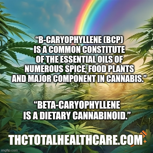
“β-caryophyllene, a bicyclic sesquiterpene widely abundant in various plant essential oils, has garnered growing attention for its potential biological effects and therapeutic benefits in liver diseases. This review systematically evaluates preclinical evidence on the pharmacological properties of BCP with emphasis on its hepatoprotective effects primarily through its anti-inflammatory, antioxidant, antifibrotic, and immunomodulatory actions.
BCP is classified as a dietary cannabinoid due to its ability to activate cannabinoid type 2 receptors in the endocannabinoid system and thereby influence key cellular signaling pathways involved in lipid metabolism and tissue remodeling. Emerging studies also highlight BCP interaction with PPAR nuclear receptor and AMPK signaling, further corroborating its role in regulating lipid homeostasis.
In the present review, we compile, summarize, and critically analyze findings from in vitro and in vivo studies on nonalcoholic fatty liver disease, recently termed as metabolic dysfunction-associated fatty liver disease (MAFLD), alcoholic liver disease, and liver fibrosis, highlighting the pharmacological and molecular mechanisms underlying therapeutic effects. These studies consistently demonstrate a reduction in hepatic steatosis, collagen deposition, and hepatocellular markers reflecting a broad spectrum of hepatoprotective effects.
Taken together, the pharmacological properties and mechanistic insights place BCP as a promising natural compound with nutraceutical, phytopharmaceutical, or dietary supplement applications for liver diseases. Despite the robust preclinical evidence, clinical validation remains scarce. Therefore, regulatory toxicology and efficacy studies are needed to establish the therapeutic potential of BCP in liver diseases and its integration as a nutraceutical or phytopharmaceutical in the clinical usage.”
“BCP is one of the important constituents in Cannabis with an abundance of 35%. In addition to its presence in Cannabis, BCP is largely present in numerous edible plants.”
“In conclusion, BCP represents a promising therapeutic avenue for managing liver diseases due to its ability to modulate multiple interrelated molecular and cellular pathways.”
“With continued research, BCP has the potential to evolve from a natural product with hepatoprotective properties to an effective adjunct or alternative in liver disease therapy, offering new hope for patients and advancing the field of liver health management.”
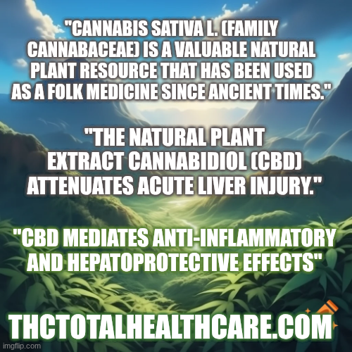
“Ethnopharmacological relevance: Acute liver injury (A-LI) is a clinical syndrome that can rapidly progress to acute liver failure, resulting in high mortality and poor prognosis. Cannabis sativa L. is an important herbaceous plant that has been widely used in folk medicine since ancient times. Cannabidiol (CBD) is its most abundant non-psychoactive compound, exhibiting hepatoprotective, anti-inflammatory, and antioxidant properties. However, the protective effect of CBD against A-LI and its mechanism remain unclear.
Objective: This study aimed to investigate the protective effects of CBD on A-LI and elucidate the underlying molecular mechanisms.
Methods: In vivo, an A-LI mouse model was induced by LPS/D-GalN. Each group was treated with or without LPS/D-GalN or CBD. H&E staining, alanine aminotransferase (ALT), aspartate aminotransferase (AST) level assay, TUNEL staining, TEM, IF, RT-qPCR, Western blot, Co-IP and adeno-associated virus (AAV) infection were performed. In vitro, RAW264.7 cells were stimulated with LPS. CCK-8, ELISA, MMP, mitochondrial ROS assay, siRNA knockdown and plasmid overexpression were performed.
Results: CBD (2.5 or 5 mg kg-1) mitigated LPS/D-GalN-induced liver damage, suppressed inflammatory cytokine expression, reduced hepatocellular apoptosis, and inhibited oxidative stress. CBD treatment increased hepatic mitofusin-2 (MFN2) protein while decreasing Parkin-MFN2 binding and MFN2 ubiquitination. In RAW264.7 cells, CBD pretreatment (2.5 or 5 μM) dose-dependently attenuated LPS-induced inflammation, apoptosis, and mitochondrial dysfunction and likewise elevated MFN2 levels while limiting its ubiquitination. MFN2 knockdown abolished CBD’s protective effects, whereas MFN2 overexpression restored them. Consistently, AAV-mediated delivery of MFN2-targeting short hairpin RNA reversed the hepatoprotective action of CBD in vivo.
Conclusion: CBD mediates anti-inflammatory and hepatoprotective effects by inhibiting MFN2 degradation through disrupting the interaction between Parkin and MFN2. These results provide molecular evidence for application of CBD in treatment of A-LI and provide references to the drug development for A-LI.”
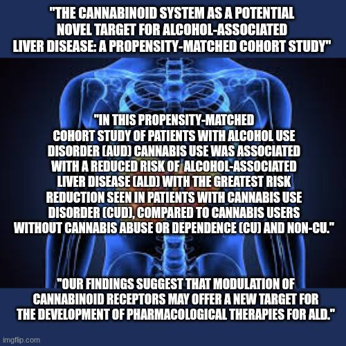
“Background: Alcohol-associated liver disease (ALD) is a leading cause of liver-related morbidity and mortality, yet effective therapeutic options remain limited. Preclinical data suggest that modulation of the hepatic endocannabinoid system, particularly via cannabidiol (CBD), may reduce alcohol-induced liver injury. Due to CBD’s limited clinical use, we sought to evaluate the association between cannabis use and ALD risk among patients with alcohol use disorder (AUD).
Methods: Using the TriNetX US Collaborative Network, we identified adult patients with AUD between 2010 and 2022. Three cohorts were constructed: cannabis use disorder (CUD), cannabis users without cannabis abuse or dependence (CU) and non-cannabis users (non-CU). Outcomes included ALD, hepatic decompensation and composite all-cause mortality over 3 years. Incidence and hazard ratios were calculated using Kaplan-Meier analysis and Cox regression.
Results: After matching, 33 114 patients were included in each of the CUD and non-CU groups. Compared to non-CU, CUD was associated with a lower risk of ALD (HR 0.60, 95% CI 0.53-0.67; p < 0.001), hepatic decompensation (HR 0.83, 95% CI 0.73-0.95; p =0.005) and all-cause mortality (HR 0.86, 95% CI 0.80-0.94; p < 0.001) among individuals with AUD. Although CU was associated with lower risks of ALD, its risks of hepatic decompensation and all-cause mortality were similar to those of the non-CU cohort with AUD.
Conclusion: In this propensity-matched cohort study of patients with AUD, cannabis use was associated with a reduced risk of ALD, with the greatest risk reduction seen in patients with CUD compared to CU and non-CU. Our findings suggest that modulation of cannabinoid receptors may offer a new target for the development of pharmacological therapies for ALD.”
“Background: Non-alcoholic fatty liver disease (NAFLD) is a common liver disorder caused by oxidative stress and dysregulation of lipid metabolism. The endocannabinoid system (ECS), particularly the type 1 cannabinoid (CB1) receptor, plays a crucial role in NAFLD progression. Cannabinoids, such as cannabidiol (CBD) and tetrahydrocannabinol (THC), along with terpenes, such as beta-myrcene and d-limonene, have shown potential therapeutic effects on liver health, particularly in reducing oxidative stress and modulating lipid metabolism.
This study aimed to analyse the effects of five cannabis oils (COs), each with different CBD:THC ratios and terpenes content, on hypertension, dyslipidemia, hepatic steatosis, oxidative stress, and CB1 receptor expression in an experimental model of NAFLD induced by a sucrose-rich diet (SRD) in Wistar rats for 3 weeks.
Methods: Male Wistar rats were fed either a: (1) reference diet (RD; standard commercial laboratory diet) or a: (2) sucrose-rich diet (SRD) for 3 weeks. 3 to 7 SRD + CO as following: (3) SRD + THC; (4) SRD + CBD; (5) SRD + CBD:THC 1:1; (6) SRD + CBD:THC 2:1; and (7) SRD + CBD:THC 3:1. The COs were administered orally at a dose of 1.5 mg total cannabinoids/kg body weight daily. The cannabinoid and terpenes content of all COs used in the study was determined. The terpenes found in COs were beta-myrcene, d-limonene, terpinolene, linalool, beta-caryophyllene, alpha-humulene, (-)-guaiol, (-)-alpha-bisabolol. During the experimental period, body weight, food intake and blood pressure were measured. Serum glucose, triglyceride, total cholesterol, uric acid, alanine aminotransferase (ALT), aspartate aminotransferase (AST), and alkaline phosphatase (AP) levels were evaluated. Liver tissue histology, NAFLD activity score (NAS), triglyceride and cholesterol content, lipogenic enzyme activities, enzyme related to mitochondrial fatty acid oxidation, reactive oxygen species (ROS), thiobarbituric acid reactive substance (TBARS), and antioxidant enzyme activities were also evaluated. The CB1 receptor expression was also determined.
Results: The results showed that SRD-fed rats developed hypertension, dyslipidemia, liver damage, hepatic steatosis, lipid peroxidation, and oxidative stress. This was accompanied by upregulation of liver CB1 receptor expression. CBD-rich CO, CBD:THC 1:1 ratio CO; CBD:THC 2:1 ratio CO and CBD:THC 3:1 ratio CO showed antihypertensive properties. THC-rich CO, CBD:THC 1:1 ratio CO; CBD:THC 2:1 ratio CO showed the greatest beneficial effects against hepatic steatosis and liver damage. All COs exhibited antioxidant effects in liver tissue. This was associated with normal liver CB1 receptor expression.
Conclusions: This study demonstrated that COs, particularly THC-rich CO, CBD:THC ratio 1:1 CO, CBD:THC ratio 2:1 CO and terpenes, can effectively reduce dyslipidemia, liver damage and hepatic steatosis in SRD-induced NAFLD. COs with a higher proportion of CBD in their composition showed antihypertensive properties. All the COs exhibited antioxidant properties. These findings suggest that COs, especially those with CBD:THC ratios of 1:1 and 2:1 and terpenes, may represent a promising therapeutic approach for managing NAFLD and preventing its progression to more severe liver disease.”
“This study demonstrated that COs, particularly THC-rich formulations, and those with CBD:THC ratios of 1:1 and 2:1, effectively reduced dyslipidemia, hepatic steatosis, and liver damage in SRD-induced NAFLD. All COs exhibited significant antioxidant properties, which contributed to the attenuation of oxidative stress. Notably, oils containing CBD also displayed antihypertensive effects, likely due to their vasodilatory properties. The modulation of CB1 receptor is closely linked to the improvement in hepatic steatosis and oxidative stress. These results underscore the synergistic role of cannabinoids and terpenes in targeting key mechanisms involved in NAFLD pathophysiology.”
“These findings suggest that COs, especially those with balanced CBD: THC ratios (1:1 and 2:1) and with meaningful terpenes content, represent a promising therapeutic approach for managing NAFLD and preventing its progression to more severe liver diseases.”
“Cannabinoids are biologically active substances acting via feedback-coupled CB1 and CB2 receptors. Their expression in myofibroblasts and liver endothelial cells is reported to be elevated in chronic liver diseases. The effect of CB1 receptor stimulation is to increase fibrosis and inflammatory activity in the liver by stimulating stellate cells, while activation of the CB2 receptor results in inhibition of fibrosis. Stimulation of the CB1 receptor may also lead to progression of liver steatosis and carcinogenesis. In end-stage liver disease, the endocannabinoid system plays an important role in the pathogenesis of encephalopathy and vascular effects, such as portal hypertension, splanchnic vasodilatation and cirrhotic cardiomyopathy. It seems that interference in endocannabinoid transmission may serve as an attractive target for the development of hepatological drugs.”
“Background: Stress-induced liver injury, resulting from acute or chronic stress, is associated with oxidative stress and inflammation. The endocannabinoid system, particularly cannabinoid receptor 2 (CB2R), plays a crucial role in liver damage. However, there are currently no clinical drugs targeting CB2R for liver diseases. Cannabidiol (CBD), a CB2R agonist, possesses anti-inflammatory and antioxidant properties. This study aims to investigate the pharmacological effects of CBD in a mouse model of stress-induced liver injury.
Methods: We employed a mouse model of stress-induced liver injury to evaluate the protective effects of CBD. Assessments included histopathological analysis, cytokine detection via ELISA, protein expression analysis using immunohistochemistry and Western blot, and gene transcription differential analysis. Transmission electron microscopy was utilized to observe mitochondrial morphology. Additionally, we examined the expression levels of CB2R, SLC7A11, α-SMA, and ACSL4 proteins to elucidate the mechanisms underlying CBD’s effects.
Results: CBD exhibited significant protective effects against stress-induced liver injury in mice. Decreases in liver function indicators (including Aspartate Aminotransferase (AST) and Alanine Aminotransferase (ALT)) and inflammatory cytokines (such as IL-1β and Tumor Necrosis Factor-alpha (TNF-α)) were observed. CBD enhanced CB2R expression and reduced α-SMA levels, mitigating liver fibrosis. It also decreased ACSL4 levels, increased SOD and GSH-Px activities, and upregulated SLC7A11 protein expression. Furthermore, CBD improved mitochondrial morphology, indicating a reduction in oxidative cell death.
Conclusion: CBD activates the CB2R/α-SMA pathway to modulate liver inflammation and fibrosis. Through the SLC7A11/ACSL4 signaling pathway, CBD alleviates oxidative stress in stress-induced liver injury, enhances mitochondrial morphology, and reduces liver damage. These findings provide a theoretical basis for the potential application of CBD in the prevention and treatment of stress-induced liver injury.”
“The cold-water immersion restraint method effectively simulates a stress-induced liver injury model caused by conditions such as hunger, cold exposure, and the fear of death. CBD demonstrates protective effects against stress-induced liver injury, and its protective mechanism may be associated with the activation of CB2R and mitochondrial metabolism. Specifically, CBD appears to exert its anti-liver fibrosis and antioxidative effects by activating CB2R, inhibiting the expression of α-SMA and ACSL4 proteins, and enhancing the expression of SLC7A11 protein, thereby alleviating liver damage.”