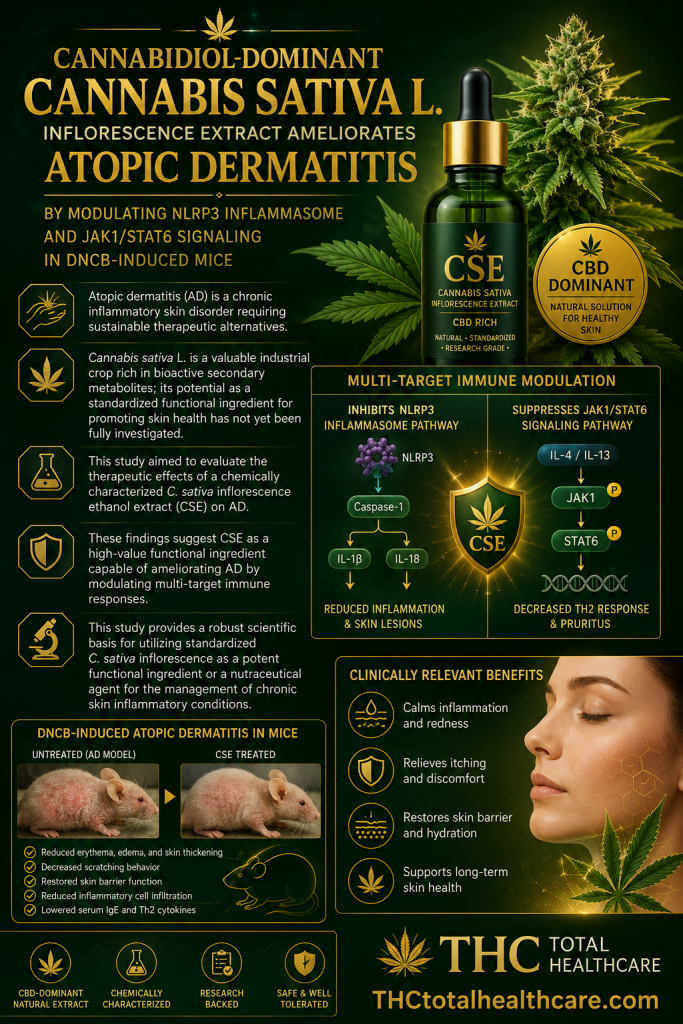
Cannabidiol hinders lipopolysaccharide-induced neutrophils migration to the lungs through suppressing nuclear factor kappa-B signal and expression of interleukin-1 beta in macrophages

Background: Acute lung injury and its more severe form, acute respiratory distress syndrome, are life-threatening diseases characterized by uncontrolled pulmonary inflammation, impaired gas exchange, and high mortality rates. Effective therapeutic agents remain limited. As a non-addictive component derived from hemp seed, the anti-inflammatory activity of cannabidiol (CBD) has been suggested by multiple pathological models.

Purpose: The purpose of this study is to investigate the potent anti-inflammatory effects of CBD in lipopolysaccharide-induced pulmonary inflammation and the mechanisms involved herein.

Methods: Mice were treated with lipopolysaccharide (LPS) intranasally to construct pulmonary inflammation model while CBD was administrated intraperitoneally at 25 mg/kg, 50 mg/kg, and 100 mg/kg. The percentage of immune cell subsets and the concentration of cytokines and chemokines were assayed to evaluate the inflammatory status of the lungs. The molecular expression of whole lungs and macrophages was obtained through RNA sequencing.

Results: The number of interstitial macrophages and neutrophils in lungs responded to the progression of inflammation and the anti-inflammatory function of CBD. In line with this, the transcriptome of lung tissue upregulated innate immune cell-related features and nuclear factor kappa-B signaling which was downregulated by CBD treatment at 50 mg/kg. CBD at this dose reduced the expression of interleukin-1 beta in both interstitial and alveolar macrophages and suppressed the expression of vascular cell adhesion molecule 1 in endothelial cells. During these processes, the mediation of inflammation was potentially conducted by interstitial macrophages.

Conclusion: CBD at 50 mg/kg significantly attenuates LPS-induced pulmonary inflammation and markedly suppresses the LPS-induced elevation in the number of neutrophils and interstitial macrophages in the lung. CBD could directly inhibit the expression of vascular cell adhesion molecule 1 in pulmonary endothelial cells and indirectly inhibit it by suppressing interleukin-1 beta secretion from macrophages, thereby reducing neutrophil infiltration into the lung and alleviating lung injury. These findings uncover the molecular mechanism whereby CBD alleviates inflammation via inhibiting granulocyte trafficking to the lungs, providing novel insights into the therapeutic potential of this compound.”

https://pubmed.ncbi.nlm.nih.gov/41935460

“Cannabidiol (CBD) is the non-addictive component in hemp seeds, known for its effects in treating constipation, reducing inflammation and pain, and providing antioxidant benefits. The anti-inflammatory properties of CBD have been well documented across diverse inflammatory disease models, with growing research interest in its therapeutic potential for pulmonary conditions”

https://www.sciencedirect.com/science/article/abs/pii/S0944711326003570?via%3Dihub

The Therapeutic Crossroad Between Mitochondria and Cannabidiol: A Mini-Review

“Cannabidiol is a non-psychoactive compound originating from Cannabis sativa L., with a promising therapeutic profile that influences numerous cellular processes. A major area of interest is its impact on mitochondria, organelles essential for cellular metabolism, ATP production, calcium homeostasis, and stress response.

This article explores the available data on contribution of CBD effect on mitochondria to its therapeutic potential in treatment of various pathologies: cancer, cardiovascular, lung, neurological, gastrointestinal and liver disease, and muscle pathologies.

Regarding cancer, the cytotoxic effects of cannabidiol on glioma, leukaemia, non-Hodgkin lymphoma, prostate, gastric, and breast cancer are analysed. In the case of cardiomyopathies and heart failure, cannabidiol plays an important role in reducing oxidative stress and promoting mitochondrial biogenesis. In lung diseases, cannabidiol reduces the expression of mitochondrial fission genes and increases the expression of fusion genes.

When it comes to neurological pathologies, cannabidiol protects neurons and exhibits a strong antioxidant effect, while in gastrointestinal and liver diseases, cannabidiol stabilises mitochondrial membrane potential, increases ATP production, and reduces oxidative stress. In muscle affections, cannabidiol improves mitochondrial function by inhibiting excessive mitophagy. Although modern formulations may improve the low bioavailability of CBD, its potential non-selective cytotoxicity toward non-malignant cells remains an important concern that warrants further investigation.

Nevertheless, cannabidiol possesses a remarkable therapeutic potential, and its effects on mitochondria open new perspectives in the treatment of numerous diseases.”

https://pubmed.ncbi.nlm.nih.gov/41892270

“In conclusion, CBD represents a molecule with remarkable therapeutic potential, and its targeting to mitochondria opens new perspectives in the treatment of chronic and degenerative diseases.”

https://www.mdpi.com/2079-7737/15/6/510

Efficacy of Cannabidiol in Reducing Virulence of Listeria monocytogenes

Listeria monocytogenes (LM) is a major foodborne pathogen causing illnesses ranging from gastroenteritis to severe systemic infections. The key virulence factors include bacterial motility, hemolysin and lecithinase production, and invasion of host tissues.

This study investigated the anti-virulence effects of cannabidiol (CBD), the main non-psychoactive compound in Cannabis sativa, against LM.

The minimum inhibitory concentration (MIC, 2289 μM; 719.8 µg/mL) and sub-inhibitory concentration (SIC, 11.92 μM; 3.75 µg/mL) of CBD were determined for LM strains Scott A and ATCC 19115. Cultures were treated with SIC, 6× SIC, 1/4× MIC, and MIC to assess effects on motility, hemolysin and lecithinase production, and adhesion and invasion of human intestinal (Caco-2) and brain endothelial (HBMEC) cells, alongside virulence gene expression by RT-qPCR. Cannabidiol’s efficacy was also determined using a Galleria mellonella larval infection model at SIC and 6× SIC.

Cannabidiol at 6× SIC significantly reduced motility, toxin production, and host cell adhesion and invasion (p < 0.05). RT-qPCR revealed downregulation of key virulence genes, including prfAhlyplcAplcBiapmotAmotBactAinlA, and inlB. In vivo, CBD enhanced larval survival in a dose-dependent manner and cytotoxicity was observed at concentrations above 33.75 µg/mL.

These results indicate that CBD, at non-bactericidal levels, effectively suppresses multiple virulence mechanisms in LM, highlighting its potential as a novel anti-virulence agent for food safety and therapeutic applications.”

https://pubmed.ncbi.nlm.nih.gov/41898547

“Cannabidiol has been reported to exert antibacterial activity through multiple, primarily membrane-associated mechanisms.”

“Collectively, these findings suggest that CBD holds promise as a prophylactic or therapeutic agent, or as an adjunct to conventional antibiotics, in mitigating listeriosis.”

https://www.mdpi.com/1422-0067/27/6/2682

Insights Into Cannabis and Cannabinoids: Chemical Properties, Legal Perspectives, and Therapeutic Applications

“Cannabis sativa L. has been used for thousands of years in various cultural, medical, and industrial settings. This review brings together evidence from historical records, plant chemical studies, clinical trials, and laws to explain the chemical properties, healing potential, and regulatory environment of cannabis and its components.

We look at over 500 identified compounds, including cannabinoids (CBs), terpenes, flavonoids, and alkaloids, along with their effects on health. The therapeutic areas covered include chronic pain, epilepsy, cancer, mental health issues, and inflammation. We also address side effects, interactions with other drugs, and approved CB-based medications.

Despite the various healing effects, gaps still exist in our understanding of the best dosing, long-term safety, and standardized product formulations. This review highlights current research directions and emphasizes the need for thorough randomized controlled trials to support the evidence-based use of cannabis in modern medicine.”

https://pubmed.ncbi.nlm.nih.gov/41822997

https://onlinelibrary.wiley.com/doi/10.1002/cbdv.202503030

Inhibitory effects of Δ8-tetrahydrocannabinol on nicotine metabolism and implications as a smoking cessation agent

“Tobacco use remains the leading cause of preventable death worldwide. The major metabolic pathway for nicotine, the addictive component in tobacco, is via cytochrome P450 (CYP) 2A6-mediated metabolism to cotinine.

Cannabidiol has been shown to reduce cigarette consumption in vivo and inhibit CYP2A6-mediated nicotine metabolism in vitro. In the present study, Δ-8-tetrahydrocannabinol (Δ8-THC), an isomer of Δ-9-tetrahydrocannabinol, was examined as a potential inhibitor of CYP2A6-mediated nicotine metabolism.

While Δ-9-tetrahydrocannabinol showed no significant inhibition of nicotine metabolism to cotinine, Δ8-THC demonstrated unbound IC50 values of 0.57 ± 0.04 μM in microsomes from recombinant wild-type CYP2A6 overexpressing human embryonic kidney 293 cells and 0.70 ± 0.16 μM in human liver microsomes (HLMs). A similar unbound IC50 value was observed for recombinant CYP2A6∗5 microsomes (0.52 ± 0.17 μM) and was modestly elevated in recombinant CYP2A6∗2 microsomes (1.00 ± 0.12 μM). IC50 shift experiments were consistent across pooled HLM (5.3-fold) and microsomes from liver specimens exhibiting the CYP2A6 (∗2/∗2) and (∗9/∗9) genotypes (6.1- and 4.0-fold, respectively) but were reduced in CYP2A6 (∗35/∗35) microsomes (1.0-fold). Irreversible inhibition kinetics in pooled HLMs by Δ8-THC yielded a kinact value of 0.022 ± 0.001 min-1 and an unbound KI value of 0.232 ± 0.062 μM. Static modeling predicted that oral dosing with 10 mg Δ8-THC increased the nicotine plasma area under the curve by 189%, with further increases observed at 20 mg and 40 mg; interactions were also observed with inhalation doses ≥70 mg.

These findings suggest that, based on CYP2A6 genotype, Δ8-THC could be a candidate for smoking cessation therapy.

SIGNIFICANCE STATEMENT: This study is the first, to the best of our knowledge, to identify Δ-8-tetrahydrocannabinol as a potent and irreversible inhibitor of nicotine metabolism to cotinine. The extent of inhibition is modulated by genetic variation in cytochrome P450 2A6. These findings suggest that further investigations focusing on Δ-8-tetrahydrocannabinol and its potential as a candidate for smoking cessation therapy are warranted.”

https://pubmed.ncbi.nlm.nih.gov/41830876

“In conclusion, the present study is, to our knowledge, the first to demonstrate the irreversible inhibition of nicotine metabolism by Δ8-THC in vitro, highlighting its potential as a smoking cessation agent.”

https://dmd.aspetjournals.org/article/S0090-9556(26)00004-8/fulltext

Effects of Cannabidiol on TAFAZZIN-Deficient B-Lymphoblastoid Cells

“Barth Syndrome (BTHS) is a debilitating X-linked genetic disorder caused by mutations in the gene encoding TAFAZZIN, an enzyme responsible for the remodeling of cardiolipin. While cyclic neutropenia is a well-recognized immunological feature of this disease, emerging evidence suggests that lymphopenia may also occur.

The objective of this study was to examine the effects of cannabidiol (CBD) on growth, cardiolipin content, and mitochondrial abnormalities in BTHS patient-derived B-lymphoblastoid cells.

CBD (1 μM) restored the growth of BTHS B-lymphoblastoids to healthy control levels, but did not alter cell cycle distribution or sub-G1 cell populations, which surprisingly also did not differ from healthy control B-lymphoblastoids. CBD treatment also fully restored the total cellular cardiolipin concentration and reversed the elevation in monolysocardiolipin/cardiolipin ratio in BTHS B-lymphoblastoids to healthy cell levels, but did not restore the cardiolipin fatty acyl composition.

Assessment of mitochondrial markers suggested that increased cardiolipin did not result from increased mitochondrial content. This improvement in cardiolipin concentration was associated with a significant increase in the maximal coupled state III respiration of BTHS B-lymphoblastoids, with all five tested BTHS donors exhibiting increased mitochondrial membrane potential following CBD treatment. CBD fully reversed the deficit in succinate dehydrogenase subunit A in BTHS cells, and partially reversed deficits in cytochrome c oxidase subunits I and IV, and partially restored supercomplex I/III2 levels, but did not rescue I/III2/IV levels.

This work suggested a potential role for CBD as a therapeutic in BTHS B-lymphopenia that merits further investigation.”

https://pubmed.ncbi.nlm.nih.gov/41823370

https://faseb.onlinelibrary.wiley.com/doi/10.1096/fj.202503384R

Driving by frequent cannabis users ‘the morning after’ last use of smoked cannabis: an observational driving simulator study

“It is well-established that cannabis can affect driving in the hours after cannabis use, but the exact duration of these effects, and relationship with delta-9-tetrahydrocannabinol (THC) concentrations in blood and oral fluid, remains to be determined.

Methods

Frequent (≥ 4 times a week) users of smoked cannabis drove a simulator the morning after (12-15 hours) last use of smoked cannabis; a control group of non-cannabis users matched for age and sex was also included. Concentrations of THC, cannabidiol (CBD) and metabolites were measured in oral fluid and blood at the time of the drive.

Results

In total, 65 participants (mean age 30 years; 33 males) in each group completed all study procedures. Participants were generally well-matched (age, sex, driving experience, amount of driving per year/week, hours of sleep) but differed in racial breakdown and years of education. Under both standard and dual task (distacted) conditions, standard deviation of lateral position (SDLP) was higher in the control group (standard: 0.305 meters; dual task: 0.272 meters; n=65) compared to the cannabis group (standard: 0.28 meters; dual task: 0.256 meters; n=65); these differences were small (Cohen’s d -0.389 (standard) and -0.359 (dual task)) and were not significant after correction for multiple comparisons. Measures of speed and following distance were not impacted. Neither blood nor oral fluid THC, CBD or metabolites was significantly correlated with any measure of driving after correction for multiple comparisons; mean concentrations of blood THC was above 2 ng/mL. After correction for multiple comparisons, trends between driving and concentrations of the psychoactive metabolite 11-hydroxy-THC (11-OH-THC) were found. Participants who smoked cannabis the night before reported higher levels of subjective intoxication, and more willingness to drive before the drive, that was not significant after correction for multiple comparisons.

Conclusions

The regular cannabis use group showed no significant impairment in driving performance 12-15 hours after last cannabis use the night before, compared to the control group. Blood and oral fluid THC concentrations may not be an accurate correlate of driving behavior. Large-scale studies are needed to determine whether less frequent users are impaired the morning after last use, and whether the present findings also extend to different routes of administration.”

https://link.springer.com/article/10.1186/s42238-026-00416-w

The effect of hemp product consumption on blood fatty acid profiles and cardiovascular disease risk factors: results of a randomized, double-blind, crossover clinical trial

“Hemp seeds are high in polyunsaturated fatty acids (PUFAs) including gamma linolenic acid (GLA), stearidonic acid (SDA), alpha linolenic acid (ALA) and linoleic acid (LA). To date, limited evidence is available on hemp product consumption and particularly hemp seeds and oil in humans and its relation to cardiometabolic risk factors.

The objective of present study was to examine the effects of hemp product consumption versus similar controls on circulating fatty acid profiles and cardiovascular disease (CVD) risk factors.

A randomized, double-blinded, crossover trial with 30 normoglycemic adults (18-65 years) within a BMI range of 25-35 kg m-2 were included. Participants consumed both hemp products and controlled products over the course of 4 weeks each. As expected, ALA (18:3 n-3), GLA (18:3 n-6) and dihomo-γ-linolenic acid (DGLA, 20:3 n-6) were elevated after the hemp treatment than controls. Similarly, ALA, DGLA as well as eicosapentaenoic acid (EPA) levels were elevated after the hemp treatment than controls. No differences in serum lipid levels, glucose and insulin concentrations, blood pressure, or body composition were observed between treatments.

Overall, consumption of hemp products modulated plasma and RBC fatty acids levels in a way which reflected the fatty acids these products are enriched in, without showing differences in major cardiometabolic risk factors. The present study demonstrated the human fatty acids profile response to consuming hemp products, novel functional foods rich in polyunsaturated fatty acids.”

https://pubmed.ncbi.nlm.nih.gov/41782552

“Overall, the present study showed that 4-week consumption of hulled hempseed and hemp oil in overweight individuals increased ALA, GLA, DGLA and EPA relative percentages in plasma and RBC respectively, demonstrating effective incorporation of hemp-derived polyunsaturated fatty acids into long-term lipid pools. These changes occurred without adverse effects on lipid metabolism, vascular function, and/or body composition.

Collectively, these findings support the metabolic safety of hemp products and highlight their potential utility as dietary sources of polyunsaturated fatty acids for improving circulating fatty acid profiles.”

https://pubs.rsc.org/en/content/articlelanding/2026/fo/d5fo04672f

Hemp-Derived Extracellular Vesicles: A Novel Frontier in Nanomedicine and Therapeutics

“While mammalian-derived extracellular vesicles (EVs) face significant challenges in clinical translation due to scalability, cost, and safety, plant-derived EVs (PDEVs) have emerged as a promising alternative.

This review focuses on EVs derived from hemp (Cannabis sativa L.), or HEVs, a particularly compelling source that combines the general benefits of PDEVs, such as improved safety and scalability, with a unique, inherent therapeutic cargo.

HEVs are naturally enriched with a potent mix of cannabinoids, terpenes, and flavonoids, which may enhance therapeutic outcomes through synergistic interactions-a phenomenon known as the ‘entourage effect.’

Preclinical studies already demonstrate their potential, showing significant anti-cancer effects against aggressive tumors like glioblastoma, along with neuroprotective and anti-inflammatory properties.

However, the critical challenge hindering their clinical application is the lack of standardized, GMP (Good Manufacturing Practice)-compliant manufacturing protocols to address the inherent biochemical variability of the source material.

Overcoming these obstacles will be vital to unlocking the potential of HEVs as a novel, scalable frontier in nanomedicine.”

https://pubmed.ncbi.nlm.nih.gov/41787227

https://link.springer.com/article/10.1007/s40259-026-00766-0

Bioactivity and Regenerative Potential of Cannabidiol in Human Dental Pulp Stem Cells: A Scoping Review of In Vitro Studies

Introduction: Cannabidiol (CBD), a nonpsychoactive compound derived from Cannabis sativa, has shown potential to influence cellular processes that are important for dental tissue repair. The aim of this scoping review was to map in vitro studies evaluating the influence of CBD on the osteogenic/odontogenic differentiation of human dental pulp stem cells (hDPSCs) in order to contribute to a better understanding of its therapeutic potential.

Methods: The review followed the Arksey and O’Malley framework, supported by the JBI Manual and PRISMA-ScR guidelines. The protocol was registered on OSF (osf.io/zfhca/). Comprehensive searches were conducted from January to June 2025 in PubMed, EMBASE, BVS, Scopus, Web of Science, ScienceDirect, and SciELO. Only studies published in English were included.

Results: Thirty articles were identified, and three in vitro studies met the eligibility criteria. At low concentrations (0.1-5 μM), CBD improved hDPSC viability, proliferation, migration, and differentiation. CBD also activated the mitogen-activated protein kinase (MAPK) and wingless-related integration site/beta-catenin signaling (WNT/β-catenin) pathways and increased the expression of odontogenic markers such as Sialophosphoprotein (DSPP), Runt-related transcription Factor 2 (RUNX2), and osteocalcin.

Conclusion: CBD shows promise as a bioactive molecule in regenerative endodontics, supporting mineralization, regulating inflammatory mediators, and promoting critical cellular activities in hDPSCs. Nevertheless, the available evidence is limited and further preclinical and clinical studies are essential to develop therapeutic protocols and assess long-term safety.

These preliminary findings indicate CBD as a novel candidate for regenerative strategies in endodontics.”

https://pubmed.ncbi.nlm.nih.gov/41767216