“G-protein coupled receptors constitute the largest family of membrane receptors and they participate in the maintenance of the homeostasis in the body. Some of these receptors still remain orphan receptors as there is insufficient research and ambiguous evidence concerning their function and endogenous ligands.
For a long time, GPR18 belonged to this group, but recently it has been classified as an endocannabinoid receptor due to its affinity to cannabinoid ligands.
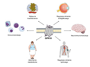
GPR18 receptor is expressed in the encephalon, thyroid gland, leukocytes, lungs and testicles. The modulatory role of GPR18 receptor has been proven in the regulation of intraocular pressure, neuroimmunomodulation, regulation of arterial blood pressure and in metabolic disorders.
In this article we summarize the current knowledge concerning the GPR18 receptor – its expression, ligands and the in the physiological processes and the pathophysiological conditions.”
“Nonalcoholic fatty liver disease (NAFLD) is a major cause of chronic liver abnormalities and has been linked with metabolic syndrome hallmarks. Unfortunately, current treatments are limited.
This work aimed to elucidate the effects of three cannabis extracts on metabolic alteration and gut microbiota composition in a mouse model of NAFLD and obesity.
Male mice were fed with a high-fat diet (HFD) for 12 weeks. Following the establishment of obesity, the HFD-fed group was subdivided into HFD or HFD that was supplemented with one of three cannabis extracts (CN1, CN2, and CN6) for additional 8 weeks. Metabolic parameters together with intestinal microbiota composition were evaluated.
Except for several minor changes in gene expression, no profound metabolic effect was found due to cannabis extracts addition. Nevertheless, marked changes were observed in gut microbiota diversity and composition, with CN1 and CN6 exhibiting microbial abundance patterns that are associated with more beneficial outcomes.
Taken together, specific cannabis extracts’ addition to an HFD results in more favorable modifications in gut microbiota. Although no marked metabolic effect was disclosed, longer treatments duration and/or higher extracts concentrations may be needed. More research is required to ascertain this conjecture and to establish the influence of various cannabis extracts on host health in general and NAFLD in particular.”
“Obesity is an abnormal or excessive accumulation of fat in the body that exacerbates metabolic and inflammatory processes, and impairs the health of afflicted individuals.
β-caryophyllene is a natural sesquiterpene that is a dietary cannabinoid with anti-inflammatory properties and potential activity against metabolic diseases.
In the present study, we evaluated the effect of β-caryophyllene on C57BL/6 mice using a diet-induced obesity model. Male mice were randomly assigned to the following groups over a 16-week period: (1) standard diet as lean control, (2) high-fat diet (HFD) as obese control, and (3) HFD + β-caryophyllene with β-caryophyllene at 50 mg/kg.
Treatment with β-caryophyllene improved various metabolic parameters including increased total body weight, fasting glucose levels, oral-glucose tolerance, insulin tolerance, fasting triglycerides, adipocyte hypertrophy, and liver macrovesicular steatosis. β-caryophyllene also modulated the levels and expression of immune response factors including adiponectin, leptin, insulin, interleukin-6, tumor necrosis factor-a, and Toll-like receptor-4.
Our data indicate that chronic supplementation with β-caryophyllene can improve relevant metabolic and immunological processes in obese mice.”
“β-caryophyllene (BCP) is a common constitute of the essential oils of numerous spice, food plants and major component in Cannabis.” http://www.ncbi.nlm.nih.gov/pubmed/23138934
“Parkinson’s disease (PD) is a complex, multifactorial neurodegenerative disease. The main pathological feature of PD is the loss or apoptosis of dopaminergic neurons in the substantia nigra (SN).
This study aimed to investigate the protective effect of cannabidiol (CBD) on 1-methyl-4-phenyl-1,2,3,6-tetrahydropyridine (MPTP)-induced neuronal dopamine injury by inhibiting neuroinflammation, which was one of the factors that cause neuronal apoptosis. Male SPF C57BL/6 mice were used to create a PD model by administering MPTP intraperitoneally for seven days and treated by oral administration of CBD for 14 days.
Behaviorally, CBD improved cognitive dysfunction and increased the number of spontaneous locomotion in PD mice. Biochemically, CBD increased the levels of 5-HT, DA and IL-10, and decreased the contents of TNF-α, IL-1β and IL-6. Pathologically, CBD increased the expression of tyrosine hydroxylase (TH). Mechanistically, CBD up-regulated the expression of Bcl-2, down-regulated the levels of Bax and Caspase-3, and repressed the expression of NLRP3/caspase-1/IL-1β inflammasome pathway.
In summary, CBD has a therapeutic effect on MPTP-induced PD mice by inhibiting the apoptosis of dopaminergic neurons and neuroinflammation. Therefore, CBD is a potential candidate for PD therapy.”
“Background: An oral route of administration for tetrahydrocannabinol (Δ9-THC) and cannabidiol (CBD) eliminates the harmful effects of smoking and has potential for efficacious cannabis delivery for therapeutic and recreational applications. We investigated the pharmacokinetics of CBD, Δ9-THC, 11-OH-THC, and 11-nor-9-carboxy-Δ9-THC (THC-COOH) in a novel oral delivery system, Solutech™, compared to medium-chain triglyceride-diluted cannabis oil (MCT-oil) in a healthy population.
Materials and Methods: Thirty-two participants were randomized and divided into two study arms employing a comparator-controlled, parallel-study design. To evaluate the pharmacokinetics of Δ9-THC, CBD, 11-OH-THC, and THC-COOH, blood was collected at pre-dose (t=0) and 10, 20, 30, and 45, min and 1, 1.5, 2, 2.5, 3, 4, 5, 6, 8, 12, 24, and 48 h post-dose after a single dose of Solutech (10.0 mg Δ9-THC, 9.76 mg CBD) or MCT (10.0 mg Δ9-THC, 9.92 mg CBD). Heart rate and blood pressure were measured at 0.5, 1, 2, 4, 6, 8, 12, 24, and 48 h. Relationships between cannabis use history, body mass index, sex, and pharmacokinetic parameters were investigated. Safety was assessed before and at 48 h post-acute dose.
Results: Acute consumption of Solutech provided a significantly greater maximum concentration (Cmax), larger elimination and absorption rate constants, faster time to Cmax and lag time, and half-life for all analytes compared to MCT-oil (p<0.001). In addition, cannabis use history had a significant influence on the pharmacokinetic parameters of CBD, Δ9-THC, 11-OH-THC, and THC-COOH. On average, participants with later age of first use had higher Δ9-THC, CBD, and THC-COOH Cmax and later time-to-Cmax and half-life for Δ9-THC, CBD, THC-COOH, and 11-OH-THC than those with earlier age of first use (p≤0.032). Those with more years of recreational cannabis use had higher area under the curve for Δ9-THC and CBD, Cmax for CBD, and longer 11-OH-THC half-life than those with less (p≤0.048).
Conclusion: This study demonstrated that consumption of Solutech enhanced most pharmacokinetics parameters measured compared to MCT-oil. Participant’s cannabis use history, including their age of first use and number of years using cannabis significantly impacted pharmacokinetic parameters investigated. Acute consumption of both products was found to be safe and well tolerated. The results suggest that Solutech may optimize bioavailability from cannabis formulations.”
“Objectives: In recent years, cannabinoids have been shown to have beneficial effects on diabetic vascular complications.
Vascular complications due to fructose-induced hyperinsulinemia (HI) and diabetic vascular complications have similar mechanisms.
The aim of this experimental study was to observe whether the cannabinoid agonist delta-9-tetrahydrocannabinol (THC) has an ameliorating effect on fructose-induced HI and vascular responses in the aortic ringof rats with HI.
Methods: A total of 24 rats were categorized into 4 groups: control (standard food pellets and water), HI (water containing 10% fructose provided for 12 weeks), THC (1.5 mg/kg/day intraperitoneal administration for 4 weeks), and THC+HI.Body weight was measured again on the last day of the study and the serum insulin level was measured with an enzyme-linked immunosorbent assay. The acetylcholine (ACh) maximum relaxant effect in aortic rings pre-contractedwith noradrenaline (NA) was evaluated.
Results: The body weight of THC and THC+HI groups was lower compared with that of the controls (p<0.01). Increasedinsulin level as a result of fructose consumption decreased with THC administration (p<0.01) while the glucose level increased in all other groups compared with the control group (p<0.01, p<0.05). The NA Emax value decreased in thegroup receiving THC treatment (p<0.01). The increased ACh pD2 value in the HI groups also decreased in the THCtreatment group (p<0.0001). The decreased maximum inhibition value in the HI group increased significantly with THC administration (p<0.001).
Conclusion: THC demonstrated beneficial effects on fructose-induced HI. THC improved ACh-induced endothelialdependent relaxation in HI rat aortic rings.”
“Objectives: A large amount of fructose is metabolized in the liver and causes hepatic functional damage. Δ9 -tetrahydrocannabinol (THC) is known as a therapeutic agent for clinical and experimental applications. The study aims to investigate the effects of THC treatment on inflammation, lipid profiles and oxidative stress in rat liver with hyperinsulinemia.
Methods: Sprague-Dawley rats were divided into groups: control, fructose (10% fructose in drinking water for 12 weeks), THC (1.5 mg/kg/day for the last 4 weeks, intraperitoneally) and fructose+THC groups. Biochemical parameters were measured spectrophotometrically. ELISA method was used for insulin measurement. Apoptosis and inflammation markers were detected by the streptavidin-biotin peroxidase method.
Key findings: The consumptions of food and fluid are inversely proportional to fructose and non-fructose groups. Insulin levels were the highest in fructose group. The reduced glutathione-S-transferase level significantly increased in fructose + THC group compared with fructose group. Total cholesterol level in the fructose + THC group was higher than the fructose group. Caspase-3 and NF-κβ immunopositive cell numbers increased in fructose + THC rats compared with fructose group. The number of IL-6 immunopositive cell decreased in fructose + THC group compared with fructose group.
Conclusions: According to the result, long-term and low-dose THC administration may reduce hyperinsulinemia and inflammation in rats to some extent.”
“Taken together, the findings from this study demonstrate that fructose consumption induces hyperinsulinemia, oxidative stress and inflammation in rats. The long-term and low-dose THC administration may prevent hyperinsulinemia and inflammation to some extent.”
“Δ9-tetrahydrocannabinol (Δ9-THC) is known for its antitumor activity and palliative effects.
However, its unfavorable physicochemical and biopharmaceutical properties, including low bioavailability, psychotropic side effects and resistance mechanisms associated to dosing make mandatory the development of successful drug delivery systems.
In this work, transferring (Tf) surface-modified Δ9-THC-loaded poly(lactide-co-glycolic) nanoparticles (Tf-THC-PLGA NPs) were proposed and evaluated as novel THC-based anticancer therapy. Furthermore, in order to assess the interaction of both the nanocarrier and the loaded drug with cancer cells, a double-fluorescent strategy was applied, including the chemical conjugation of a dye to the nanoparticle polymer along with the encapsulation of either a lipophilic or a hydrophilic dye.
Tf-THC PLGA NPs exerted a cell viability decreased down to 17% vs. 88% of plain nanoparticles, while their internalization was significantly slower than plain nanoparticles. Uptake studies in the presence of inhibitors indicated that the nanoparticles were internalized through cholesterol-associated and clathrin-mediated mechanisms.
Overall, Tf-modification of PLGA NPs showed to be a highly promising approach for Δ9-THC-based antitumor therapies, potentially maximizing the amount of drug released in a sustained manner at the surface of cells bearing cannabinoid receptors.”
“The potential therapeutic applications of marijuana, firstly reported in 1997 by the National Institutes of Health (NIH, USA), are attributed to a great extent to its main component, Δ9-tetrahydrocannabinol (Δ9-THC)1. This cannabinoid continues to attract special attention in oncology due to its palliative effects and antitumor activity; Δ9-THC has been reported to inhibit tumor angiogenesis and cell growth in malignant tissues, leading to cell death.”
“Δ9-THC has been reported to inhibit tumor angiogenesis and cell growth in malignant tissues.”
“Overall, Tf-modification of PLGA NPs seemed a highly promising approach for Δ9-THC-based antitumor therapies, aiming at a prolonged action of the carrier at the target cell surface. Moreover, the translation of this strategy to the delivery of alternative active pharmaceutical ingredients with pharmacological targets on the surface of cells could lead to advances in related therapies.”
“There is evidence that demonstrates the effect of cannabinoid agonists inhibiting relevant aspects in lung cancer, such as proliferation or epithelial-to-mesenchymal transition (EMT).
Most of these studies are based on evidence observed in in vitro models developed on cancer cell lines. These studies do not consider the complexity of the tumor microenvironment (TME). One of the main components of the TME is cancer-associated fibroblasts (CAFs), cells that are relevant in the control of proliferation and metastasis in lung cancer.
In this work, we evaluated the direct effects of two cannabinoid agonists, tetrahydrocannabinol (THC) and cannabidiol (CBD), used alone or in combination, on CAFs and non-tumor normal fibroblasts (NFs) isolated from adenocarcinoma or from healthy lung tissue from the same patients.
We observed that these compounds decrease cell density in vitro and inhibit the increase in the relative expression of type 1 collagen (COL1A1) and fibroblast-specific protein 1 (FSP1) induced by transforming growth factor beta (TGFβ). On the other hand, we studied whether THC and CBD could modulate the interactions between CAFs or NFs and cancer cells. We conditioned the culture medium with stromal cells treated or not with THC and/or CBD and cultured A549 cells with them.
We found that culture media conditioned with CAFs or NFs increased cell density, induced morphological changes consistent with EMT, inhibited cadherin-1 (CDH1) gene expression, and induced an increase in the relative expression of cadherin-2 (CDH2) and vimentin (VIM) genes in A549 cells. These changes were inhibited or decreased by THC and CBD administered alone or in combination. In another series of experiments, we conditioned culture media with A549 cells treated or not with THC and/or CBD, in the presence or absence of TGFβ. We observed that culture media conditioned with A549 in the presence of TGFβ induced an increase in the expression of COL1A1 and VIM, both in CAFs and in non-tumor NFs. Both THC and CBD ameliorated these effects.
In summary, the results presented here reinforce the usefulness of cannabinoid agonists for the treatment of some relevant aspects of lung cancer pathology, and demonstrate in a novel way their possible effects on CAFs as a result of their relationship with cancer cells. Likewise, the results reinforce the usefulness of the combined use of THC and CBD, which has important advantages in relation to the possibility of using lower doses, thus minimizing the psychoactive effects of THC.”
“The medical application of cannabidiol (CBD) has been gathering increasing attention in recent years. This non-psychotropic cannabis-derived compound possesses antiepileptic, antipsychotic, anti-inflammatory and anxiolytic properties. Recent studies report that it also exerts antineoplastic effects in multiple types of cancers, including melanoma.
In this in vitro study we tried to reveal the anticancer properties of CBD in malignant melanoma cell lines (SK-MEL 28, A375, FM55P and FM55M2) administered alone, as well as in combination with mitoxantrone (MTX) or cisplatin (CDDP).
The effects of CBD on the viability of melanoma cells were measured by the MTT assay; cytotoxicity was determined in the LDH test and proliferation in the BrdU test. Moreover, the safety of CBD was tested in human keratinocytes (HaCaT) in LDH and MTT tests.
Results indicate that CBD reduces the viability and proliferation of melanoma-malignant cells and exerts additive interactions with MTX. Unfortunately, CBD produced antagonistic interaction when combined with CDDP. CBD does not cause significant cytotoxicity in HaCaT cell line.
In conclusion, CBD may be considered as a part of melanoma multi-drug therapy when combined with MTX. A special attention should be paid to the combination of CBD with CDDP due to the antagonistic interaction observed in the studied malignant melanoma cell lines.”