Antioxidant Activity of Hemp ( Cannabis sativa L.) Seed Oil in Drosophila melanogaster Larvae under Non-Stress and H 2 O 2-Induced Oxidative Stress Conditions

antioxidants-logo

“The oil extracted from hemp seeds has significant nutritional and biological properties due to the unique composition of polyunsaturated fatty acids and various antioxidant compounds. The potential of this oil for the prevention of oxidative stress and for the treatment of oxidative-stress-induced ailments is of increasing interest. Most studies of hemp seed oil were conducted in-vitro, meaning we lack information about effects and activity in vivo. In the present study, we evaluated the hypothesis that hemp seed oil at different concentrations improves the oxidative state of D. melanogaster, under non-stress as well as hydrogen-peroxide-induced stress. We analyzed the effects of hemp seed oil on oxidative stress markers and on the life cycle of D.melanogaster under non-stress and hydrogen-peroxide-induced stress conditions. D.melanogaster larvae were exposed to hemp seed oil concentrations ranging from 12.5 to 125 μL/mL. The results revealed that under non-stress conditions, oil concentrations up to 62.5 µL/mL did not induce negative effects on the life cycle of D. melanogaster and maintained the redox status of the larval cells at similar levels to the control level. Under oxidative stress conditions, biochemical parameters were significantly affected and only two oil concentrations, 18.7 and 31.2 µL/mL, provided protection against hydrogen peroxide stress effects. A higher oil concentration (125 μL/mL) exerted negative effects on the oxidative status and increased larval mortality. The tested oil was characterized chemically by NMR, transesterification, and silylation, followed by GC-MS analyses, and was shown to contain polyunsaturated fatty acid triglycerides and low levels of tocopherols. The high levels of linoleic and linolenic acids in the oil are suggested to be responsible for the observed in vivo antioxidant effects. Taken together, the results show that hemp seed oil is effective for reducing oxidative stress at the cellular level, thus supporting the hypothesis. The obtained results point to the potential of hemp seed oil for the prevention and treatment of conditions caused by the action of reactive oxygen species.”

https://pubmed.ncbi.nlm.nih.gov/34067432/

https://www.mdpi.com/2076-3921/10/6/830

A review on the techno-functional, biological, and health-promoting properties of hempseed-derived proteins and peptides

Journal of Food Biochemistry

“Protein-energy malnutrition is a global challenge that demands urgent attention, especially with the increasing population growth and unmatched food security plans. One strategy is to expand the list of protein sources, such as neglected and underutilized crops, with high protein content. A good number of plant proteins, in addition to their nutritional benefits, exert therapeutic properties as seen in seeds derived from legumes and emerging sources such as hemp. In this review, the transepithelial transport, functional, and biological properties of hempseed proteins (HSPs) and peptides were discussed. The review also described the potential safety issues of incorporating hempseeds in food products. Due to the multitargeted effects of hempseed-derived proteins and their peptides against many chronic diseases, and their functional properties, current knowledge shows that hempseed has tremendous potential for functional food and nutraceutical applications.

PRACTICAL APPLICATIONS: The alarming rate of malnutrition and the attendant health consequences demand that underexploited nutrient-rich crops should be incorporated as part of our common dietary sources. Among these crops, hempseed is gaining attention as an emerging source of proteins and peptides with promising potential in prevention and management of chronic diseases such as diabetes, hypertension, cancer, hypercholesterolemia, obesity, and diseases whose etiology involves oxidative stress and inflammation. Fortunately, a growing body of research evidence is demonstrating that hempseed is a reservoir of proteins and peptides with nutraceutical potentials for curbing life-threatening diseases.”

https://pubmed.ncbi.nlm.nih.gov/35312074/

https://onlinelibrary.wiley.com/doi/10.1111/jfbc.14127


Tobacco, but Neither Cannabis Smoking Nor Co-Drug Use, Is Associated With Hearing Loss in the National Health and Nutrition Examination Survey, 2011 to 2012 and 2015 to 2016

Ear and Hearing - Home | Facebook


“Introduction:
A relationship between tobacco smoking and hearing loss has been reported; associations with cannabis smoking are unknown. In this cross-sectional population-based study, we examined relationships between hearing loss and smoking (tobacco, cannabis, or co-drug use).

Methods: We explored the relationship between hearing loss and smoking among 2705 participants [mean age = 39.41 (SE: 0.36) years] in the National Health and Nutrition Examination Survey (2011 to 12; 2015 to 16). Smoking status was obtained via questionnaire; four mutually exclusive groups were defined: nonsmokers, current regular cannabis smokers, current regular tobacco smokers, and co-drug users. Hearing sensitivity (0.5 to 8 kHz) was assessed, and two puretone averages (PTAs) computed: low- (PTA0.5,1,2) and high-frequency (PTA3,4,6,8). We defined hearing loss as threshold >15 dB HL. Multivariable logistic regression was used to examine sex-specific associations between smoking and hearing loss in the poorer ear (selected based on PTA0.5,1,2) adjusting for age, sex, race/ethnicity, hypertension, diabetes, education, and noise exposure with sample weights applied.

Results: In the age-sex adjusted model, tobacco smokers had increased odds of low- and high-frequency hearing loss compared with non-smokers [odds ratio (OR) = 1.58, 95% confidence ratio (CI): 1.05 to 2.37 and OR = 1.97, 95% CI: 1.58 to 2.45, respectively]. Co-drug users also had greater odds of low- and high-frequency hearing loss [OR = 2.07, 95% CI: 1.10 to 3.91 and OR = 2.24, 95% CI: 1.27 to 3.96, respectively]. In the fully adjusted multivariable model, compared with non-smokers, tobacco smokers had greater odds of high-frequency hearing loss [multivariable adjusted odds ratio = 1.64, 95% CI: 1.28-2.09]. However, in the fully adjusted model, there were no statistically significant relationships between hearing loss (PTA0.5,1,2 or PTA3,4,6,8) and cannabis smoking or co-drug use.

Discussion: Cannabis smoking without concomitant tobacco consumption is not associated with hearing loss. However, sole use of cannabis was relatively rare and the prevalence of hearing loss in this population was low, limiting generalizability of the results. This study suggests that tobacco smoking may be a risk factor for hearing loss but does not support an association between hearing loss and cannabis smoking. More definitive evidence could be derived using physiological measures of auditory function in smokers and from longitudinal studies.”

https://pubmed.ncbi.nlm.nih.gov/35383601/

https://journals.lww.com/ear-hearing/Abstract/9900/Tobacco,_but_Neither_Cannabis_Smoking_Nor_Co_Drug.3.aspx

Management of chronic pain with Jalaprakshalana (water-wash) Shodhita (processed) Bhanga ( Cannabis sativa L.) in cancer patients with deprived quality of life: An open-label single arm clinical trial

“Introduction: Pain is a common and complex symptom of cancer having physical, social, spiritual and psychological aspects. Approximately 70%-80% of cancer patients experiences pain, as reported in India. Ayurveda recommends use of Shodhita (Processed) Bhanga (Cannabis) for the management of pain but no research yet carried out on its clinical effectiveness.

Objective: To assess the analgesic potential of Jala-Prakshalana (Water-wash) processed Cannabis sativa L. leaves powder in cancer patients with deprived quality of life (QOL) through openlabel single arm clinical trial.

Materials and methods: Waterwash processed Cannabis leaves powder filled in capsule, was administered in 24 cancer patients with deprived QOL presenting complaints of pain, anxiety or depression; for a period of 4 weeks; in a dose of 250 mg thrice a day; along with 50 ml of cow’s milk and 4 g of crystal sugar. Primary outcome i.e. pain was measured by Wong-Bakers FACES Pain Scale (FACES), Objective Pain Assessment (OPA) scale and Neuropathic Pain Scale (NPS). Secondary outcome namely anxiety was quantified by Hospital Anxiety and Depression Scale (HADS), QOL by FACT-G scale, performance score by Eastern Cooperative Oncology Group (ECOG) and Karnofsky score.

Results: Significant reduction in pain was found on FACES Pain Scale (P < 0.05), OPA (P < 0.05), NPS (P < 0.001), HADS (P < 0.001), FACT-G scale (P < 0.001), performance status score like ECOG (P < 0.05) and Karnofsky score (P < 0.01).

Conclusion: Jalaprakshalana Shodhita Bhanga powder in a dose of 250 mg thrice per day; relieves cancerinduced pain, anxiety and depression significantly and does not cause any major adverse effect and withdrawal symptoms during trial period.”

https://pubmed.ncbi.nlm.nih.gov/31831967/

“Administration of Jalaprakshalana Shodhita Bhanga (water-wash processed Cannabis) leaves powder in dose of 250 mg thrice a day with 50 ml of cow’s milk and 4 g sugar as an adjuvant, for a period of 1 month; significantly relieves pain, anxiety and depression of cancer patients without creating any major side effects, dependency and withdrawal symptoms. Processed Cannabis is significantly effective for improvement in QOL of a cancer patient.”

https://www.ayujournal.org/article.asp?issn=0974-8520;year=2019;volume=40;issue=1;spage=34;epage=43;aulast=Tavhare

Heavy and Chronic Cannabis Addiction does not Impact Motor Function: BOLD-fMRI Study

Generic placeholder image

“Objective: The aim of this paper is to demonstrate the impact of heavy and chronic cannabis use on brain potential functional control, reorganization, and plasticity in the cortical area.

Methods: 23 cannabis users were convened in 3 user’s groups. The first group included 11 volunteers with an average of 15 joins/day; the second group included 6 volunteers with an average of 1.5 joins/day; the third group included 6 volunteers with an average of 2.8 joins/week. Besides, a 6 healthy volunteers (control group). All healthy and cannabis users underwent identical brain BOLD-fMRI assessment of the motor function. Besides, neuropsychological and full biological assessments were achieved.

Results: BOLD-fMRI maps of motor areas were obtained, including quantitative evaluation of the activations in the motor area. Besides, statistical analysis of various groups was achieved.

Conclusion: Chronic cannabis addiction of varying use strength by groups of heavy, moderate, low dose, and zero doses are shown to have systematically equivalent effects on the control of brain motor function. Indeed, the BOLD-fMRI shows a remarkable sensitivity to minimal brain plasticity and reorganization of the functional motor control of the studied cortical area, and such varionation was not shown. Specific elucidation of the cannabis effect mechanisms in this unique function should clarify further protective pharmacological effects. This might illuminate the use of neuronal resources to prepare processes for pharmacological use and pharmaceutical forms. This suggests exploring any potential cannabis pharmaceutical form in diseases involving motor impairments.”

https://pubmed.ncbi.nlm.nih.gov/35578884/

https://www.eurekaselect.com/article/123583

Indeterminacy of cannabis impairment and ∆ 9-tetrahydrocannabinol (∆ 9-THC) levels in blood and breath

Scientific Reports

“Previous investigators have found no clear relationship between specific blood concentrations of ∆9-tetrahydrocannabinol (∆9-THC) and impairment, and thus no scientific justification for use of legal “per se” ∆9-THC blood concentration limits. Analyzing blood from 30 subjects showed ∆9-THC concentrations that exceeded 5 ng/mL in 16 of the 30 subjects following a 12-h period of abstinence in the absence of any impairment. In blood and exhaled breath samples collected from a group of 34 subjects at baseline prior to smoking, increasing breath ∆9-THC levels were correlated with increasing blood levels (P < 0.0001) in the absence of impairment, suggesting that single measurements of ∆9-THC in breath, as in blood, are not related to impairment. When post-smoking duration of impairment was compared to baseline ∆9-THC blood concentrations, subjects with the highest baseline ∆9-THC levels tended to have the shortest duration of impairment. It was further shown that subjects with the shortest duration of impairment also had the lowest incidence of horizontal gaze nystagmus at 3 h post-smoking compared to subjects with the longest duration of impairment (P < 0.05). Finally, analysis of breath samples from a group of 44 subjects revealed the presence of transient cannabinoids such as cannabigerol, cannabichromene, and ∆9-tetrahydrocannabivarin during the peak impairment window, suggesting that these compounds may be key indicators of recent cannabis use through inhalation. In conclusion, these results provide further evidence that single measurements of ∆9-THC in blood, and now in exhaled breath, do not correlate with impairment following inhalation, and that other cannabinoids may be key indicators of recent cannabis inhalation.”

https://pubmed.ncbi.nlm.nih.gov/35585089/

“In conclusion, we present further evidence that single measurements of ∆9-THC in blood cannot establish impairment, that single measurements of ∆9-THC in exhaled breath likewise do not correlate with impairment, and that ∆9-THCV and CBC may be key indicators of recent cannabis use through inhalation within the impairment window.”

https://www.nature.com/articles/s41598-022-11481-5

Comparative study of CNR1 and CNR2 cannabinoid receptors expression levels in COVID-19 patients with and without diabetes mellitus: Recommendations for future research targets

Diabetes & Metabolic Syndrome: Clinical Research & Reviews

“Background and aims: The COVID-19 pandemic has prompted researchers to look for effective therapeutic targets. The effect of endocannabinoid system against infectious diseases is investigated for several years. In this study, we evaluated the expression level of CNR1 and CNR2 genes in patients with COVID-19 with and without diabetes to provide new insights regarding these receptors and their potential effect in COVID-19 disease.

Methods: In this study, peripheral blood monocytes cells (PBMCs) were isolated from eight different groups including COVID-19 patients, diabetic patients, and healthy individuals. RNA were extracted to evaluate the expression level of CNR1 and CNR2 genes using real-time PCR. The correlation between the expression levels of these genes in different groups were assessed.

Results: A total of 80 samples were divided into 8 groups, with each group consisting of ten samples. When comparing severe and moderate COVID-19 groups to healthy control group, the expression levels of the CNR1 and CNR2 genes were significantly higher in the severe and moderate COVID-19 groups. There were no significant differences between the mild COVID-19 group and the healthy control group. It was found that the expression levels of these genes in patients with diabetes who were infected with SARS-COV-2 did not differ across COVID-19 groups with varying severity, but they were significantly higher when compared to healthy controls.

Conclusion: Our study suggests the possible role of endocannabinoid system during SARS-COV-2 pathogenicity as the expression of CNR1 and CNR2 were elevated during the disease.”

https://pubmed.ncbi.nlm.nih.gov/35580523/

“In conclusion, the outcomes of this research supports the possible role of endocannabinoid system during SARS-COV-2 pathogenicity as the expression of CNR1 and CNR2 were elevated during the disease. Moreover, despite their limitations due to psychiatric side effects, the regulated use of cannabinoids should be examined by researchers to identify their potential effectiveness as a therapeutic target in COVID-19 disease.”

https://www.sciencedirect.com/science/article/pii/S1871402122001138?via%3Dihub

A Review of Hemp as Food and Nutritional Supplement

View details for Cannabis and Cannabinoid Research cover image

“The term “hemp” refers to Cannabis sativa cultivars grown for industrial purposes that are characterized by lower levels of tetrahydrocannabinol (THC), the active principle responsible for Cannabis psychotropic effects. Hemp is an extraordinary crop, with enormous social and economic value, since it can be used to produce food, textiles, clothing, biodegradable plastics, paper, paint, biofuel, and animal feed, as well as lighting oil.

Various parts of the hemp plant represent a valuable source of food and ingredients for nutritional supplements. While hemp inflorescence is rich in nonpsychoactive, yet biologically active cannabinoids, such as cannabidiol (CBD), which exerts potent anxiolytic, spasmolytic, as well as anticonvulsant effects, hempseed has a pleasant nutty taste and represents a valuable source of essential amino acids and fatty acids, minerals, vitamins, and fibers. In addition, hempseed oil is a source of healthy polyunsaturated fatty acids, and hemp sprouts are rich in antioxidants.

This review article aims to provide a comprehensive outlook from a multidisciplinary perspective on the scientific evidence supporting hemp beneficial properties when consumed as food or supplement. Marketing of hemp-derived products is subjected to diversified and complex regulations worldwide for several reasons, including the fact that CBD is also the active principal of pharmaceutical agents and that regulatory bodies in some cases ban Cannabis inflorescence regardless of its THC content. Some key regulatory aspects of such a complex scenario are also analyzed and discussed in this review article.”

https://pubmed.ncbi.nlm.nih.gov/33614949/

https://www.liebertpub.com/doi/10.1089/can.2020.0001

Hemp Seeds of the Polish ‘Bialobrzeskie’ and ‘Henola’ Varieties ( Cannabis sativa L. var. sativa) as Prospective Plant Sources for Food Production

molecules-logo

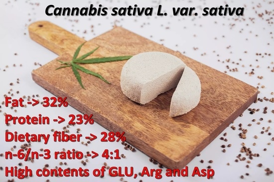
“This publication characterizes the nutritional value of the Polish hemp seeds of the ‘Bialobrzeskie’ and ‘Henola’ varieties, including the profile/content of fatty acids and amino acids. Hemp seeds were found to be rich in protein, fat, and dietary fiber. Polyunsaturated fatty acids (PUFA) dominated the unsaturated fatty acids (UFA) profile. Their average share within the total fatty acids (FA) was as high as 75%. Linoleic acid belonging to this group accounted for 55% of the total FA. Lipid profile indices (Σ n – 6/Σ n – 3, Σ PUFA/Σ SFA, the thrombogenicity index, the atherogenicity index and the hypocholesterolemic/hypercholesterolemic ratio) proved the high nutritional value of hemp oil. Considering the tyrosine + phenylalanine and histidine contents, hemp protein exhibited a great degree of similarity to egg protein, which is known and valued for its high biological value.”

https://pubmed.ncbi.nlm.nih.gov/35209234/

https://www.mdpi.com/1420-3049/27/4/1448


The Role of Cannabidiol (CBD) in a Cisplatin-Induced Model of Chronic Neuropathic Pain

“Cannabinoid-based therapies offer a safer, non-opioid alternative for the management of chronic pain. While most studies focus on the analgesic potential of the main psychoactive component of marijuana, Δ9-tetrahydrocannabinol, fewer studies have investigated the role of the non-psychoactive component, cannabidiol (CBD). CBD has been purported to have analgesic, anti-inflammatory, anticonvulsant, and anxiolytic effects. In addition to having actions at both cannabinoid receptors (CB1 and CB2 ), CBD has been shown to interact with both the transient receptor potential vanilloid-1 (TRPV1) and serotonergic (5-HT) receptors. Clinically, CBD’s lack of psychoactivity and decreased abuse liability make it an appealing pharmacotherapeutic for the management of chronic pain. Therefore, the purpose of the current study was to determine whether CBD sex- or dose-dependently reverses antinociception in an acute model of thermal pain and/or mechanical allodynia in a model of cisplatin-induced chronic neuropathic pain. Furthermore, we observed the degree to which CB1 , CB2 , 5-HT, and TRPV1 receptors may be mediating these anti-allodynic responses. Male and female wild-type mice were assessed for either the anti-allodynic effects of 0, 1, 3, 10, and 30 mg/kg CBD in a cisplatin-induced model of neuropathic pain or the antinociceptive effects of 0, 1, 3, 10, 30, and 100 mg/kg CBD in a model of acute thermal (tail-flick) pain 60 minutes following CBD administration. To determine the relative contributions of each receptor subtype in mediating the anti-allodynic effects of CBD, male and female mice were pretreated with either: vehicle, the CB1 inverse agonist SR141716A (10 mg/kg), the CB2 antagonist SR144528 (10 mg/kg), the TRPV1 antagonist capsazepine (10 mg/kg), or the 5-HT2 antagonist methysergide (4 mg/kg) 30 minutes prior to treatment with CBD. Mice were assessed for the effects of the pretreatment alone and in combination with CBD. CBD at a dose of 3 mg/kg was able to partially reverse cisplatin-induced allodynia in male and female mice, while doses of 10 and 30 mg/kg resulted in nearly complete reversal. Our preliminary findings showed that the anti-allodynic effects of 30 mg/kg CBD were completely blocked following pretreatment with SR141716A and SR144528, and partially blocked by capsazepine in both male and female mice. Interestingly, pretreatment with methysergide partially attenuated the anti-allodynic effects of CBD in females alone. In contrast, CBD (0-100 mg/kg) failed to induce antinociception on the tail-flick assay. CBD did induce mild hypothermia with males showing a greater degree of CBD-mediated hypothermia than female mice. Taken together, these findings suggest that CBD may be a more effective treatment option for the management of chronic pain. This study highlights the therapeutic potential of CBD in a model of neuropathic pain and suggests that these effects may have clinical implications for the use of cannabinoids in chronic pain management.”

https://pubmed.ncbi.nlm.nih.gov/35560789/

https://faseb.onlinelibrary.wiley.com/doi/10.1096/fasebj.2022.36.S1.R5197