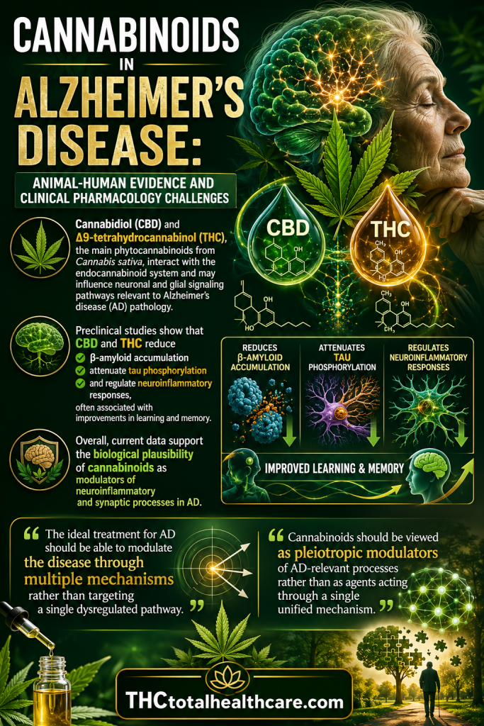
“Alzheimer’s disease (AD) is characterized by enhanced β-amyloid peptide (βA) deposition along with glial activation in senile plaques, selective neuronal loss, and cognitive deficits.
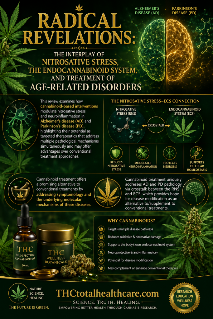
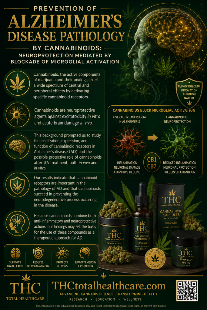
Cannabinoids are neuroprotective agents against excitotoxicity in vitro and acute brain damage in vivo.
This background prompted us to study the localization, expression, and function of cannabinoid receptors in AD and the possible protective role of cannabinoids after βA treatment, both in vivo and in vitro.
Here, we show that senile plaques in AD patients express cannabinoid receptors CB1 and CB2, together with markers of microglial activation, and that CB1-positive neurons, present in high numbers in control cases, are greatly reduced in areas of microglial activation. In pharmacological experiments, we found that G-protein coupling and CB1 receptor protein expression are markedly decreased in AD brains. Additionally, in AD brains, protein nitration is increased, and, more specifically, CB1 and CB2 proteins show enhanced nitration. Intracerebroventricular administration of the synthetic cannabinoid WIN55,212-2 to rats prevent βA-induced microglial activation, cognitive impairment, and loss of neuronal markers.
Cannabinoids (HU-210, WIN55,212-2, and JWH-133) block βA-induced activation of cultured microglial cells, as judged by mitochondrial activity, cell morphology, and tumor necrosis factor-α release; these effects are independent of the antioxidant action of cannabinoid compounds and are also exerted by a CB2-selective agonist. Moreover, cannabinoids abrogate microglia-mediated neurotoxicity after βA addition to rat cortical cocultures.
Our results indicate that cannabinoid receptors are important in the pathology of AD and that cannabinoids succeed in preventing the neurodegenerative process occurring in the disease.”
“Cannabinoid receptors in AD brain.”
“Cannabinoids, the active components of marijuana and their analogs, exert a wide spectrum of central and peripheral effects by activating specific cannabinoid receptors, two of which have been well characterized to date: CB1 and CB2.”
“Cannabinoids exert neuroprotection under different experimental conditions. Thus, cannabinoid receptor activation protects hippocampal or granule cerebellar neurons from excitotoxicity”
“This background prompted us to study the characteristics and localization of cannabinoid receptors in AD brain, with particular emphasis on any relationship with microglial activation.”
“Cannabinoid treatment prevents βA-induced microglial activation and neurotoxicity in vitro.”
“Cannabinoid treatment prevents βA-induced toxic effects in vivo.”
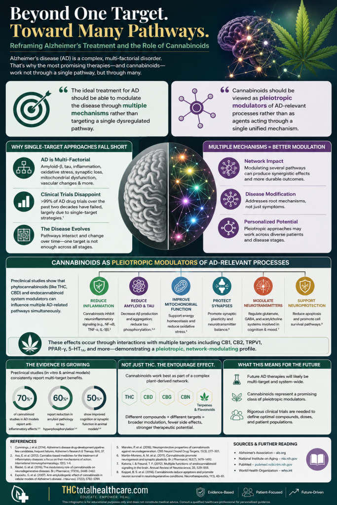
“Because cannabinoids combine both anti-inflammatory and neuroprotective actions, our findings may set the basis for the use of these compounds as a therapeutic approach for AD.”