The effects of cannabinoids in exemestane-resistant breast cancer cells: PS181.

“Exemestane is one of the aromatase inhibitors (AI) used as first line treatment for estrogen-receptor positive breast cancer in post-menopausal women. Exemestane acts by inhibiting aromatase, the enzyme responsible for the conversion of androgens to estrogens and also by promoting apoptosis of breast cancer cells. Nevertheless, despite its therapeutic success, this AI, after prolonged treatment, can induce acquired resistance, which causes tumor relapse. Therefore, it is important to find new strategies to overcome resistance in order to improve breast cancer treatment.

Considering that the development of resistance is the main reason for endocrine treatment failure, our group decided to explore the ability of three cannabinoids, Δ9-tetrahydrocannabinol (THC), cannabidiol (CBD) and anandamide (AEA), to reverse resistance to exemestane. The THC and CBD are phytocannabinoids derived from the plant Cannabis sativa (marijuana) whereas AEA is an endocannabinoid. For that, it was used LTEDaro cells, a long-term estrogen deprived ER+ breast cancer cell line that mimics resistance to exemestane. These cells were treated with exemestane in combination with two phytocannabinoids, CBD and THC, and the endocannabinoid AEA.

The presence of CB1 and CB2 in LTEDaro cells was confirmed by Western blot analysis and the effects of the combination of cannabinoids with exemestane were evaluated by MTT and LDH assays. Cell morphology was analyzed by Giemsa and Hoechst staining.

Results: Our results demonstrate that all the cannabinoids induce a decrease in viability of exemestane-resistant cells, in a dose- and time-dependent manner, without LDH release. These results indicate that the studied cannabinoids, mainly THC and AEA, revert the resistance to exemestane, probably by inducing apoptosis, as observed in Giemsa/Hoechst stain by the presence of typical morphological features of apoptosis.

Conclusion: This study highlights the efficacy of using cannabinoids as a potential adjuvant treatment to revert resistance to AIs.”

https://www.ncbi.nlm.nih.gov/pubmed/32258721

https://journals.lww.com/pbj/fulltext/2017/09000/The_effects_of_cannabinoids_in.118.aspx

n-3 Polyunsaturated Fatty Acid Amides: New Avenues in the Prevention and Treatment of Breast Cancer.

ijms-logo “Over the last decades a renewed interest in n-3 very long polyunsaturated fatty acids (PUFAs), derived mainly from fish oils in the human diet, has been observed because of their potential effects against cancer diseases, including breast carcinoma. These n-3 PUFAs mainly consist of eicosapentaenoic acid (EPA) and docosahexaenoic acid (DHA) that, alone or in combination with anticancer agents, induce cell cycle arrest, autophagy, apoptosis, and tumor growth inhibition. A large number of molecular targets of n-3 PUFAs have been identified and multiple mechanisms appear to underlie their antineoplastic activities. Evidence exists that EPA and DHA also elicit anticancer effects by the conversion to their corresponding ethanolamide derivatives in cancer cells, by binding and activation of different receptors and distinct signaling pathways. Other conjugates with serotonin or dopamine have been found to exert anti-inflammatory activities in breast tumor microenvironment, indicating the importance of these compounds as modulators of tumor epithelial/stroma interplay. The objective of this review is to provide a general overview and an update of the current n-3 PUFA derivative research and to highlight intriguing aspects of the potential therapeutic benefits of these low-toxicity compounds in breast cancer treatment and care.”

https://www.ncbi.nlm.nih.gov/pubmed/32224850

https://www.mdpi.com/1422-0067/21/7/2279

“Anticancer effects of n-3 EPA and DHA and their endocannabinoid derivatives on breast cancer cell growth and invasion.”  https://www.ncbi.nlm.nih.gov/pubmed/31679810

Inhibition of autophagic flux differently modulates cannabidiol-induced death in 2D and 3D glioblastoma cell cultures.

 Scientific Reports“Radiotherapy combined with chemotherapy is the major treatment modality for human glioblastoma multiforme (GBM). GBMs eventually relapse after treatment and the average survival of GBM patients is less than two years.

There is some evidence that cannabidiol (CBD) can induce cell death and increases the radiosensitivity of GBM by enhancing apoptosis. Beside initiation of death, CBD has been demonstrated as an inducer of autophagy.

In the present study, we address the question whether CBD simultaneously induces a protective effect in GBM by upregulating autophagy. Addition of chloroquine that suppressed autophagic flux to 2D GBM cultures increased CBD-induced cell death, presenting proof for the protective autophagy.

Blockage of autophagy upregulated radiation-induced cytotoxicity but only modestly affected the levels of cell death in CBD- or CBD/γ-irradiated 3D GBM cultures. Furthermore, CBD enhanced the pro-apoptotic activities of JNK1/2 and MAPK p38 signaling cascades while partially downregulated the pro-survival PI3K-AKT cascade, thereby changing a balance between cell death and survival.

Suppression of JNK activation partially reduced CBD-induced cell death in 3D GBM cultures. In contrast, co-treatment of CBD-targeted cells with inhibitors of PI3K-AKT-NF-κB, IKK-NF-κB or JAK2-STAT3 pathways killed surviving GBM cells in both 2D and 3D cultures, potentially improving the therapeutic ratio of GBM.”

https://www.ncbi.nlm.nih.gov/pubmed/32060308

“Killing efficiency of cannabinoids (CBD, THC and their combination CBD+THC) against GBM in vitro and in animal experiments has been elucidated in numerous studies during the last 15 years. Additional investigations also confirmed a cytotoxic role of cannabinoids for several other types of cancer. A number of studies demonstrated the efficiency of combined treatments of cannabinoids together with γ-irradiation in both cell culture and in animal experiments.”

https://www.nature.com/articles/s41598-020-59468-4

The role of cannabinoids in the treatment of cancer.

“The aim of this review article is to summarize current knowledge about the role of cannabinoids and cannabinoid receptors in tumor disease modulation and to evaluate comprehensively the use of cannabinoids in cancer patients.

METHOD:

According to the PRISMA protocol, we have included data from a total of 105 articles.

RESULTS:

Cannabinoids affect cancer progression by three mechanisms. The most important mechanism is the stimulation of autophagy and affecting the signaling pathways leading to apoptosis. The most important mechanism of this process is the accumulation of ceramide. Cannabinoids also stimulate apoptosis by mechanisms independent of autophagy. Other mechanisms by which cannabinoids affect tumor growth are inhibition of tumor angiogenesis, invasiveness, metastasis, and the modulation of the anti-tumor immune response.

CONCLUSION:

In addition to the symptomatic therapy of cancer patients, the antitumor effects of cannabinoids (whether in monotherapy or in combination with other cancer therapies) have promising potential in the treatment of cancer patients. More clinical trials are needed to demonstrate the antitumor effect of cannabinoids.”

https://www.ncbi.nlm.nih.gov/pubmed/31950844

http://www.elis.sk/index.php?page=shop.product_details&flypage=flypage.tpl&product_id=6509&category_id=158&option=com_virtuemart&vmcchk=1&Itemid=1

Cannabidiol promotes apoptosis via regulation of XIAP/Smac in gastric cancer.

Image result for cell death and disease“According to recent studies, Cannabidiol (CBD), one of the main components of Cannabis sativa, has anticancer effects in several cancers. However, the exact mechanism of CBD action is not currently understood.

Here, CBD promoted cell death in gastric cancer.

We suggest that CBD induced apoptosis by suppressing X-linked inhibitor apoptosis (XIAP), a member of the IAP protein family. CBD reduced XIAP protein levels while increasing ubiquitination of XIAP. The expression of Smac, a known inhibitor of XIAP, was found to be elevated during CBD treatment. Moreover, CBD treatment increased the interaction between XIAP and Smac by increasing Smac release from mitochondria to the cytosol. CBD has also been shown to affect mitochondrial dysfunction.

Taken together, these results suggest that CBD may have potential as a new therapeutic target in gastric cancer.”

https://www.ncbi.nlm.nih.gov/pubmed/31699976

“In conclusion, our study showed that CBD induces apoptotic cell death in gastric cancer cells, which is triggered by ER stress generation and subsequent XIAP inhibition by Smac. Taken together, our results suggest the potential of CBD in novel treatments against gastric cancer.”

 https://www.nature.com/articles/s41419-019-2001-7

figure7

Anticancer effects of n-3 EPA and DHA and their endocannabinoid derivatives on breast cancer cell growth and invasion.

Prostaglandins, Leukotrienes and Essential Fatty Acids Home“The anticancer effects of the omega-3 long chain polyunsaturated fatty acids (LCPUFA), EPA and DHA may be due, at least in part, to conversion to their respective endocannabinoid derivatives, eicosapentaenoyl-ethanolamine (EPEA) and docosahexaenoyl-ethanolamine (DHEA).

Here, the effects of EPEA and DHEA and their parent compounds, EPA and DHA, on breast cancer (BC) cell function was examined. EPEA and DHEA exhibited greater anti-cancer effects than EPA and DHA in two BC cells (MCF-7 and MDA-MB-231) whilst displaying no effect in non-malignant breast cells (MCF-10a).

Both BC lines expressed CB1/2 receptors that were responsible, at least partly, for the observed anti-proliferative effects of the omega-3 endocannabinoids as determined by receptor antagonism studies. Additionally, major signalling mechanisms elicited by these CB ligands included altered phosphorylation of p38-MAPK, JNK, and ERK proteins.

Both LCPUFAs and their endocannabinoids attenuated the expression of signal proteins in BC cells, albeit to different extents depending on cell type and lipid effectors. These signal proteins are implicated in apoptosis and attenuation of BC cell migration and invasiveness.

Furthermore, only DHA reduced in vitro MDA-MB-231 migration whereas both LCPUFAs and their endocannabinoids significantly inhibited invasiveness. This finding was consistent with reduced integrin β3 expression observed with all treatments and reduced MMP-1 and VEGF with DHA treatment.

Attenuation of cell viability, migration and invasion of malignant cells indicates a potential adjunct nutritional therapeutic use of these LCPUFAs and/or their endocannabinoids in treatment of breast cancer.”

https://www.ncbi.nlm.nih.gov/pubmed/31679810

https://www.plefa.com/article/S0952-3278(19)30112-7/fulltext

Endocannabinoid system and the expression of endogenous ceramides in human hepatocellular carcinoma.

 Journal Cover
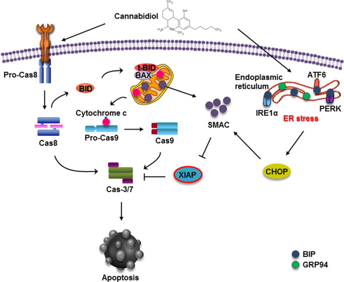
“The endogenous lipid metabolism network is associated with the occurrence and progression of malignancies.

Endocannabinoids and ceramides have demonstrated their anti-proliferative and pro-apoptotic properties in a series of cancer studies.

The aim of the present study was to evaluate the expression patterns of endocannabinoids and endogenous ceramides in 67 pairs of human hepatocellular carcinoma (HCC) tissues and non-cancerous counterpart controls.

Anandamide (AEA), the major endocannabinoid, was reduced in tumor tissues, probably due to the high expression and activity of fatty acid amide hydrolase. Another important endocannabinoid, 2-arachidonylglycerol (2-AG), was elevated in tumor tissues compared with non-tumor controls, indicating that the biosynthesis of 2-AG is faster than the degradation of 2-AG in tumor cells.

Furthermore, western blot analysis demonstrated that cannabinoid receptor 1 was downregulated, while cannabinoid receptor 2 was elevated in HCC tissues, in accordance with the alterations in the levels of AEA and 2-AG, respectively. For HCC tissues, the expression levels of C18:0, 20:0 and 24:0-ceramides decreased significantly, whereas C12:0, 16:0, 18:1 and 24:1-ceramides were upregulated, which may be associated with cannabinoid receptor activation and stearoyl-CoA desaturase protein downregulation.

The exact role of endocannabinoids and ceramides in regulating the fate of HCC cells requires further investigation.”

https://www.ncbi.nlm.nih.gov/pubmed/31423220

https://www.spandidos-publications.com/10.3892/ol.2019.10399

Antiproliferative and antioxidant effect of polar hemp extracts (Cannabis sativa L., Fedora cv.) in human colorectal cell lines.

Publication Cover “Total phenolic content and antioxidant activity of polar extracts of edible resources from Fedora hemp cultivar (Cannabis sativa L.), namely seed, flour and oil, were evaluated. The main components in the polar extracts were identified using HPLC-DAD and HPLC-ESI-MS/MS. As expected, the molecular profile of components from seeds and flour was strictly similar, dominated by N-trans-caffeoyltyramine. The profile of oil polar extracts contained hydroxycinnamic acid derivatives and cannabinoids at lower extent. While the extracts from hemp seed and flour did not interfere with growth of Caco-2 and HT-29 cell, the one from oil (150 µg/mL) significantly reduced cell viability after 24 h of treatment. This effect was associated with the activation of apoptotic cell death and was independent from the antioxidant capacity of the oil polar extract. Notably, HT-29 cells differentiated with sodium butyrate were not sensitive to the cytotoxic effect of the oil extract.”

https://www.ncbi.nlm.nih.gov/pubmed/31544542

https://www.tandfonline.com/doi/abs/10.1080/09637486.2019.1666804?journalCode=iijf20

The protective effect of cannabinoid type 2 receptor activation on renal ischemia-reperfusion injury.

“Kidney ischemia reperfusion (IR) injury is an important health problem resulting in acute renal failure. After IR, the inflammatory and apoptotic process is triggered.

The relation of Cannabinoid type 2 (CB2) receptor with inflammatory and apoptotic process has been determined. The CB2 receptor has been shown to be localized in glomeruli and tubules in human and rat kidney. Activation of CB2 receptor with JWH-133 has been shown to reduce apoptosis and inflammation.

In this study, it was investigated whether CB2 activation with selective CB2 receptor agonist JWH-133 was protective against renal IR injury.

We found that JWH-133 and CB2 receptor activation had a curative effect against kidney IR damage. JWH-133 may be a new therapeutic agent in preventing kidney IR damage.”

https://www.ncbi.nlm.nih.gov/pubmed/31446615

https://link.springer.com/article/10.1007%2Fs11010-019-03616-6

Preclinical evidence on the anticancer properties of phytocannabinoids

Image result for CROSBI“Phytocannabinoids are unique terpenophenolic compounds predominantly produced in the glandular trichomes of the cannabis plant (Cannabis sativa L.). The delta-9- tetrahydrocannabinol (THC) is the main active constituent responsible for the plant’s psychoactive effect and, together with the non- psychoactive cannabidiol (CBD), the most investigated naturally occurring cannabinoid.

The first report on the antitumor properties of cannabis compounds appeared more than forty years ago, but the potential of targeting the endocannabinoid system in cancer has recently attracted increasing interest. Our study aimed to review the last decade’s findings on the anticancer potential of plant- derived cannabinoids and the possible mechanisms of their activity.

A large body of in vitro data has been accumulated demonstrating that phytocannabinoids affect a wide spectrum of tumor cells, including gliomas, neuroblastomas, hepatocarcinoma as well as skin, prostate, breast, cervical, colon, pancreatic, lung and hematological cancer.

It has been found that they can stop the uncontrolled growth of cancer cells through the cell-cycle arrest, inhibition of cell proliferation and induction of autophagy and apoptosis. They can also block all the steps of tumor progression, including tumor cell migration, adhesion and invasion as well as angiogenesis. The observed effects are mainly mediated by the cannabinoid CB1 and/or CB2 receptors, although some other receptors and mechanisms unrelated to receptor stimulation may also be involved.

The majority of available animal studies confirmed that phytocannabinoids are capable of effectively decreasing cancer growth and metastasis in vivo. THC was found to be effective against experimental glioma, liver, pancreatic, breast and lung cancer while CBD showed activity against glioma and neuroblastoma, melanoma, colon, breast, prostate and lung cancer. Further in vitro and in vivo studies also greatly support their use in combination with traditional chemotherapy or radiotherapy, which results in improved efficiency, attenuated toxicity or reduced drug resistance.

Taken together most of available preclinical results emphasize the extensive therapeutic potential of THC and CBD in various types of cancers. The potential clinical interest of cannabinoids is additionally suggested by their selectivity for tumor cells as well as their good tolerance and the absence of normal tissue toxicity, which are still the major limitations of most conventional drugs. The accumulated preclinical evidence strongly suggests the need for clinical testing of cannabinoids in cancer patients.”